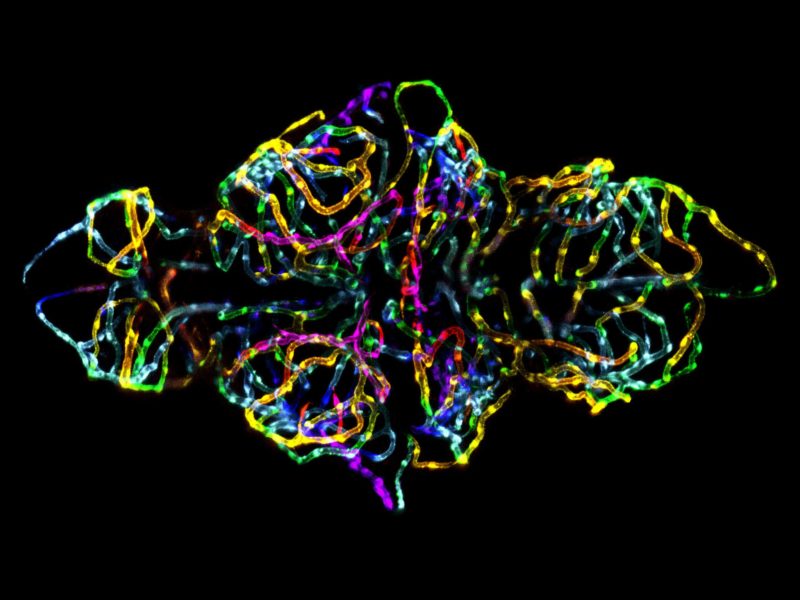
Dr. Jennifer Peters’ and Dr. Michael Taylor’s winning image of the blood-brain barrier in a live zebrafish embryo perfectly demonstrates the intersection of art and science that drives the Nikon Small World Competition.
The blood-brain barrier plays a critical role in neurological function and disease. Drs. Peters and Taylor, developed a transgenic zebrafish to visualize the development of this structure in a live specimen. By doing so, this model proves that not only can we image the blood-brain barrier, but we can also genetically and chemically dissect the signaling pathways that modulate the blood-brain barrier function and development.
To achieve this image, Peters and Taylor used a maximum intensity projection of a series of images acquired in the z plane. The images were first pseudo-colored with a rainbow palette based on depth so that the coloring scheme would be both visually appealing and provide spatial information. In doing so, Peters and Taylor captured an image that Peters says“not only captures the beauty of nature, but is also topical and biomedically relevant.”
Both Peters and Taylor have more than ten years of imaging experience. Peters is an imaging scientist in the St. Jude Children’s Research Hospital’s Light Microscopy Core Facility and Taylor is an Assistant Member in the Department of Chemical Biology and Therapeutics at St. Jude Children’s Research.