Time-Lapse Video of Zebrafish “Inner Ear” Development Wins Small World in Motion Competition
Posted on April 27, 2015
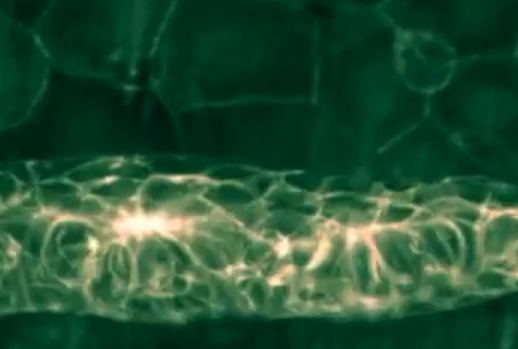
Nikon Instruments Inc. is pleased to announce the winners of the fourth annual Nikon Small World in Motion Photomicrography Competition. First place for the 2014 competition is awarded to Dr. Mariana Muzzopappa (of the Institute for Research in Biomedicine Barcelona) and Jim Swoger (of the Center for Genomic Regulation, Barcelona), for their stunning capture of the development of a zebrafish lateral line – a process that could provide insight into curing deafness in humans.
The mesmerizing first-place video demonstrates the development of the zebrafish lateral line, a sensory organ analogous to the inner ear of humans, that in fish senses movements in the surrounding water. Muzzopappa and Swoger captured this time-lapse footage by using a combination of transgenes in the fish to label undifferentiated and support cells, and track hair cell differentiation throughout a 36-hour timeframe. The result is an examination of organ development that is stunning not only in its visual effect, but the potential it represents for scientists to one-day replicate the process to counter hearing loss in humans.
Dr. Douglas Clark of Paedia Corporation took second place with a time-lapse look at crystals forming in a single drop of a saturated solution of caffeine in water. This 20-minute process is compressed into just 40 seconds in the video, revealing the beautifully chaotic formation of linear crystalline segments in a constrained space. Clark used polarized diascopic illumination to reveal the rainbow crystals that formed in the solution, a dynamic return to equilibrium from a liquid state.
Finally, Dr. John Hart, professor emeritus of the University of Colorado, Boulder, earned third place for his video of volatile oil film on a water surface. As a professor of atmospheric and oceanic sciences for many years, Hart aimed to capture the small-scale dynamics of evaporation, instability, and coalescence within the oil film that are important to the longevity of fuel spills. Hart also revealed an unexpected beauty, using reflected light differential interference contrast (DIC) techniques to capture the vibrant, almost-shimmering colors.
“The capability to capture and share the movement or development of a specimen under the microscope clearly represents one of the greater advancements in the tools available to the scientific community in recent years – and we are honored to shine a light on some of the best examples each year,” said Eric Flem, Communications and CRM manager, for Nikon Instruments. “We are continually amazed that this equipment is involved not only in doing the cutting edge science, but also enabling us all to witness it firsthand. As the deadline for this year’s competition approaches, we hope that these winning videos will inspire other scientists and science-enthusiasts to share the beauty and motion they capture under a microscope as well.”
The 2014 Nikon Small World in Motion completion was judged by Paul Maddox, Assistant Professor of Biology at the University of North Carolina Chapel Hill.
THE OFFICIAL 2014 NIKON SMALL WORLD IN MOTION WINNERS
The following are the Top Three Winners and Honorable Mentions for Nikon Small World in Motion 2014. The full gallery of winning videos can be viewed at www.nikonsmallworld.com.
1st Place
Dr. Mariana Muzzopappa with Jim Swoger
Institute for Research in Biomedicine Barcelona
Barcelona, Spain
The development of the zebrafish lateral line, the organ that senses water movements in the fish
4D SPIM (Selective Plane Illumination Microscopy)
20x/0.5
2nd Place
Dr. Douglas Clark
Paedia Corporation
San Francisco, California, USA
Time-lapse of caffeine crystallization
Polarized light
3rd Place
Dr. John Hart
Dept. Atmospheric and Oceanic Sci, Univ. Colorado
Boulder, Colorado, USA
Oil film floating on water
Reflected Light Differential Interference Contrast (Nomarski)
12.5x
HONORABLE MENTIONS
Dr. Nancy Costigliola-Tarsky
Harvard Medical School
Brookline, Massachusetts, USA
Vimentin (a protein) undergoing mitosis
Confocal
40x
Mr. Ralph Grimm
Jimboomba, Queensland, Australia
Rotifer (microscopic water creature)
Differential Interference Contrast
750x
Mr. Gerd A. Günther
Düsseldorf, Germany
Sulfur crystals
Brightfield, Polarized Light
100x
Mr. Elliott Hagedorn
Boston Children’s Hospital
Boston, Massachusetts, USA
Time-lapse of a 2 day old zebrafish embryo with green erythrocytes and red blood vessels
Confocal
Dr. Nils Lindstrom
The Roslin Institute
Edinburgh, Scotland
Developing mouse embryonic kidneys
Fluorescence
4x
Dr. Jeremy Logue
National Institutes of Health
Bethesda, Maryland, USA
Human melanoma (cancer) cells blebbing (distorting)
Confocal
100x
Ms. Xi Lu
University of Maryland
Washington, D.C., USA
Chitosan micromotors in water and hydrogen peroxide
Macroscope
2x, 6x
Mr. Pushkar Paranjpe
Post Doctoral Fellow
Bangalore, Karnataka, India
Tracking the locomotion of a fruit fly
200 Frames/s video acquired using an AVT Pike camera
10x
Dr. Masha Prager-Khoutorsky
Centre for Research in Neuroscience
Montreal, Quebec, Canada
Aplysia (sea slug) neuron
Confocal
100x
Mr. Alex Ritter with Dr. Bi-Chang Chen (Academia Sinica, Taipei, Taiwan), Dr. Wesley Legant (Janelia Farm Research Campus, Ashburn, VA), Dr. Liang Gao (Stony Brook University, Stony Brook, NY)
National Institutes of Health
Washington, D.C., USA
Killer T cell engaging a cancer cell
Light Sheet Microscopy
1000x
Ms. Ashley Smith
CFD Research Corporation
Huntsville, Alabama, USA
Leukocyte Inflammation Response
Fluorescence
10x
Mr. Wim van Egmond
Micropolitan MuseumBerkel en Rodenrijs, Netherlands
Lacrymaria olor (ciliate)
Differential Interference Contrast
160x
Mr. Shaohe Wang
University of California, San Diego
La Jolla, California, USA
C. elegans (nematode) cell division
Confocal
63x
Mr. Kevin Yehl
Emory University
Atlanta, Georgia, USA
DNA micro-machine rolling across a fluorescently labeled RNA surface
Brightfield
See the 2014 Small World in Motion winners