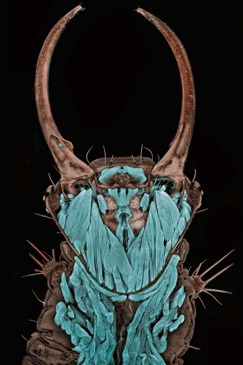
In this stunning winning image, confocal microscopy offers an intimate perspective on the invertebrate’s inner architecture, including horn-like mandibles, powerful muscles, simple eyes and a small central nervous system. A collection of multiple images stitched together allows a large area to be visualized in great detail. While the image is aesthetically interesting, it also provides insight into the complex, intricate muscles required for the seemingly simple task of operating the insect’s mouth; thus demonstrating the power and applicability of confocal microscopy to invertebrate morphology studies.
2011 Photomicrography Competition
1st Place
Portrait of a Chrysopa sp. (green lacewing) larva
Dr. Igor Robert Siwanowicz
- Affiliation
- Max Planck Institute of Neurobiology
Martinsried, Germany
- Technique
- Confocal
- Magnifaction
- 20