HaCaT mitotic cell, FISH stained for telomeres (yellow) and DAPI (gray), reconstructed in 3D
2012 Photomicrography Competition
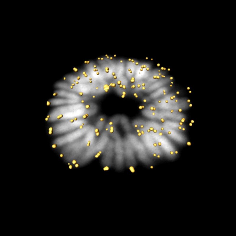
Image of Distinction
HaCaT cells in prometaphase
Damir Krunic
- Affiliation
- German Cancer Research Center
Heidelberg, Germany
- Technique
- Magnifaction
- 63