2013 Photomicrography Competition
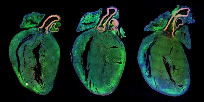
Rat heart sections

Dr. Miguel Mano

Affiliation
International Centre for Genetic Engineering and Biotechnology
Department of High-Throughput Screening
Trieste, Italy
Technique
Fluorescence
Magnification
4x

This image shows sections of rat hearts. The muscle cells of the heart, called cardiomyocytes, are stained in green, nucleus of cells are counterstained in blue and proliferating cells are labeled in green.

Mouse adipose (fat) tissue

Dr. Daniela Malide

Affiliation
National Institutes of Health (NIH)
National Heart, Lung and Blood Institute, Light Microscopy Core Facility
Bethesda, Maryland, USA
Technique
Confocal, 2-photon
Magnification
25x

Mizapin (antidepressant drug) crystals

Dr. Robert Markus

Affiliation
Stockholm University
Department of Neurochemistry
Stockholm, Sweden
Technique
Polarized Light, Differential Interference Contrast
Magnification
160x