The image shows an entire zebrafish embryo at single-cell resolution and enables an insightful view of its early morphogenetic development. The embryo’s cell nuclei were fluorescently labeled and imaged with a SiMView custom light-sheet microscope. Color encodes depth in the image.
2014 Photomicrography Competition
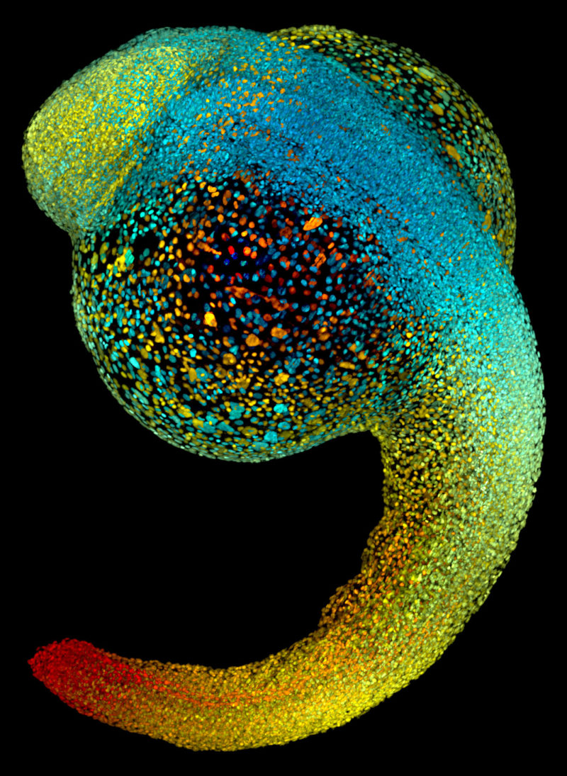
Image of Distinction
Live zebrafish embryo at 22 hours post-fertilization
Dr. Philipp Keller
- Affiliation
- Howard Hughes Medical Institute (HHMI)
Ashburn, Virginia, USA
- Technique
SiMView Light-Sheet Microscopy
- Magnifaction
- 0