This photograph shows ammonite shell under high magnification. Specimens such as this are sold for jewelry under the name amolite.
2016 Photomicrography Competition
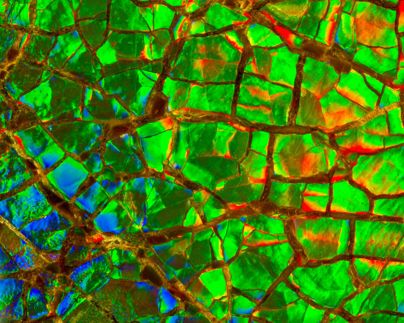
Image of Distinction
Ammonite shell (Placenticeras meeki)
Norm Barker
- Affiliation
- Johns Hopkins School of Medicine
Department of Pathology & Art as Applied to Medicine
Baltimore, Maryland, USA
- Technique
- Magnifaction
- 0