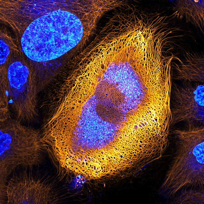
Dr. Bram van den Broek reveals an exceptional and microscopic view of something we see every day — our skin. The image depicts a cell expressing an excessive amount of keratin, seen here fluorescently labeled in yellow. He came across this peculiar but beautiful skin cell while researching the dynamics of keratin filaments with Andriy Volkov, a student in the Cell Biophysics group. The group is led by Professor Kees Jalink at The Netherlands Cancer Institute (NKI). Dr. van den Broek assists many research groups at NKI’s bio imaging facility, where he specializes in advanced fluorescence microscopy techniques and image analysis.
Keratin is an important structural protein in skin cells. The keratin fibrous network protects the cells against mechanical stress, and is involved in many other cellular functions, like cell migration and adhesion. Studying the structure, dynamics and regulation of the keratin network can reveal information about such processes. In certain types of cancer, for instance, reduced amounts of specific keratins are indicative for tumor aggressiveness.
“There are more than 50 different keratin proteins known in humans. The expression patterns of keratin are often abnormal in skin tumor cells, and it is widely used as tumor marker in diagnostics,” said. Dr. van den Broek. “By studying the ways different proteins like keratin dynamically change within a cell, we can better understand the progression of cancers and other diseases.”