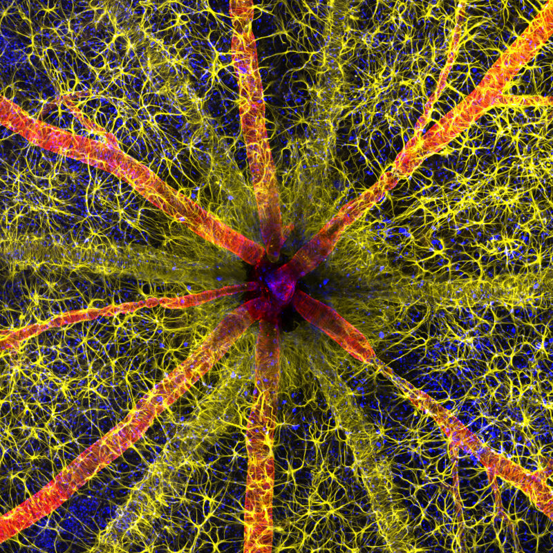
Assisted by Jayden Dickson, Hassanain Qambari won the 2023 Nikon Small World Photomicrography competition for his vivid image of a rodent optic nerve head showing astrocytes (yellow), contractile proteins (red), and retinal vasculature (green). The colorful image provides an important contribution to the study and reversal of diabetic retinopathy.
“Current diagnostic criteria and treatment regimens for diabetic retinopathy are limited to the late-stage appearance of the disease, with irreversible damage to retinal microvasculature and function,” said Qambari. “The visual system is a complex and highly specialized organ, with even relatively minor perturbations to the retinal circulation able to cause devastating vision loss. I entered the competition as a way to showcase the complexity of retinal microcirculation.”
It was no easy task to capture his winning image and at some points, a technically demanding challenge. After locating the fine vessels near 110 microns in diameter, Qambari established a protocol for labeling different cell types. “Over the past 20 years, our research group has refined the technique of isolated ocular perfusion labeling for fine vessels in the eye,” said Qambari. “While the ophthalmic artery in the rodent model presented a challenge, we were able to overcome it with persistence and patience.”