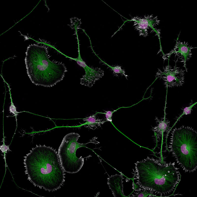
The first place prize for the 2024 Nikon Small World Photomicrography Competition was awarded to Dr. Bruno Cisterna, with assistance from Dr. Eric Vitriol of Augusta University, for his groundbreaking image of differentiated mouse brain tumor cells, highlighting the actin cytoskeleton, microtubules, and nuclei. This image reveals how disruptions in the cell's cytoskeleton – the structural framework and “highways” known as microtubules – can lead to diseases like Alzheimer's and ALS.
Dr. Cisterna’s research revealed that profilin 1 (PFN1), a protein crucial for building the cell's structure, plays a key role in maintaining the microtubule highways essential for cellular transport. When PFN1 or related processes are disrupted, these highways can malfunction, leading to cellular damage similar to what is observed in neurodegenerative diseases.
"One of the main problems with neurodegenerative diseases is that we don't fully understand what causes them,” said Dr. Cisterna. “To develop effective treatments, we need to figure out the basics first. Our research is crucial for uncovering this knowledge and ultimately finding a cure. Differentiated cells could be used to study how mutations or toxic proteins that cause Alzheimer's or ALS alter neuronal morphology, as well as to screen potential drugs or gene therapies aimed at protecting neurons or restoring their function.”
His patience and determination were crucial in capturing his image. "I spent about three months perfecting the staining process to ensure clear visibility of the cells. After allowing five days for the cells to differentiate, I had to find the right field of view where the differentiated and non-differentiated cells interacted. This took about three hours of precise observation under the microscope to capture the right moment, involving many attempts and countless hours of work to get it just right.”
The hard work behind this discovery underscores its significance, bringing researchers closer to answers that could potentially transform millions of lives. “After three years of research, we finally published our findings four months ago in the Journal of Cell Biology, and there's still more work to be done,” said Dr. Cisterna. “I’m deeply passionate about scientific imaging; I’ve been following the Nikon Small World contest for about 15 years. It's an incredible contest that highlights the beauty of photomicrography but also inspires continued exploration and innovation in the field."


 Share
Share Tweet
Tweet Pin-It