2025 Photomicrography Competition
8th Place
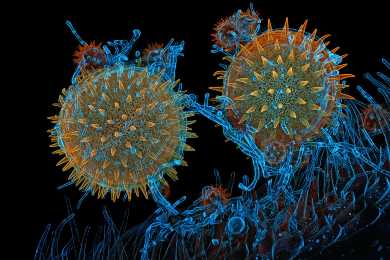
Mallow pollen germinating on stigma while being parasitized by a filamentous fungus
Dr. Igor Robert Siwanowicz
- Affiliation
- Howard Hughes Medical Institute (HHMI)
Janelia Research Campus
Ashburn, Virginia, USA
- Technique
- Confocal
- Magnifaction
- 40X (Objective Lens Magnification)
In Their Own Words
What does being a winner of the Nikon Small World competition mean to you?
It’s always an honor to place in this long-standing and prestigious contest. I’m happy to have an opportunity to bring the beauty of natural forms to the broader public’s attention; It’s a privilege to share my interests with those that appreciate them.
Can you describe in further detail (beyond what’s written in the caption), in a way the general public would understand, what is happening in your winning image?
The image shows several pollen grains resting on the stigma of a mallow flower. The larger grains are from the mallow, the smaller – from an aster flower. The larger grains are germinating – releasing their pollen tubes carrying male gametes down to the ovules contained at the base of the flower. The process of fertilization was unlikely to have had occurred in this case – the thin, segmented strands wrapping around the pollen grains and branching inside of them are the hyphae of a filamentous parasitic fungus, possibly from the genus Cladosporium or Alternaria. Mallow pollen reaches up to 150 micrometers in diameter; daisy pollen is about 20 micrometers wide.
Why do you think competitions like Nikon Small World, and scientific communication in general, are important?
For most non-experts, microscopy images, with their rather unfamiliar and abstract shapes, may cause confusion. Hopefully this is the good kind of confusion: the kind that leads to learning. It is sometimes referred to as an Optimal Confusion and it is thought to be a key epistemic emotion, since it leads to knowledge acquisition. Confusion like that causes a certain degree of discomfort: a mental itch that comes with the recognition of a gap in one’s knowledge. That itch can only be scratched by finding the information that will close that gap.
Awe and wonder are emotions often experienced in response to things like music, art, vast landscapes, the northern lights, or a lightning storm. The size of the phenomenon doesn’t need to be a defining criterium. For me, it is the exploration of the fine details and intricacies of natural forms and designs that often triggers those emotions. Awe and wonder are both particularly vital states of mind in our human experience. Both are humbling – they make us feel small, yet connected - to ourselves, others, nature, and the universe. While awe makes us stop and take it all in, wonder inspires a desire to understand, to engage with the object or phenomenon. Wonder sparks active interest and incites deeper curiosity. Perhaps unsurprisingly, the research suggests that an attitude of curiosity increases life satisfaction and rejuvenates the brain – it actually delays the onset of and slows down cognitive decline as we age, making curiosity a physically healthy emotion, certainly worthy of lifelong cultivation.
Microscopy can awaken viewers to the beauty of natural forms and the many facets of design that can be found in nature. It has power to incite interest and curiosity, and motivate some passionate investigations into the natural world.