Developing Sensory Nervous System of a Zebrafish Wins 2018 Nikon Small World in Motion Competition
Posted on September 27, 2018
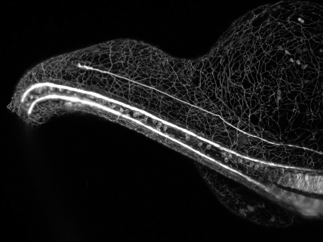
Nikon Instruments Inc. today unveiled the winners of the eighth annual Nikon Small World in Motion Photomicrography Competition. First place was awarded to Dr. Elizabeth Haynes and Jiaye "Henry" He for their video of a zebrafish embryo growing its elaborate sensory nervous system. The video reflects a time lapse of 16 hours and uses gentle light sheet technology to capture the whole zebrafish embryo in 3D, at a high temporal resolution. Dr. Haynes studies the role of kinesin light chain genes during the highly complex development of sensory neurons, while Mr. He specializes on developing microscopy technology to image living specimens at the best possible resolution.
“There are many kinesin light chain genes and their individual roles are poorly understood,” said Dr. Haynes. “If we can learn what changes in axon growth may occur when different kinesin light chain genes are perturbed, we can better understand their functions in the development of neurons, and their potential roles in neurodegenerative disorders such as Alzheimer's disease.”
To film the developing embryo, Dr. Haynes and Mr. He chose to let the zebrafish embryo grow in water, where it develops naturally inside their home-built microscope. This is a much more challenging approach as the specimen could easily move out of the field of view. The conventional technique of mounting the zebrafish in a block of gel restricts the growth of the embryo, which can impact the development of the neurons and result in a less accurate study.
“This year’s video represents exactly the kind of cutting edge scientific imaging we strive to showcase in the Nikon Small World in Motion competition,” said Eric Flem, Communications Manager, Nikon Instruments, “As microscope and imaging technologies advance, we are seeing scientifically relevant events better than ever before in visually beautiful detail.”
Dr. Haynes added, “I hope people see this video and understand how much we share with other organisms in terms of our development. A neuron is a neuron, and it’s really amazing how most of the time development goes right when so much could go wrong. There is so much art occurring within science and nature, and it’s really special to watch.”
This year’s second place winner moves from the biological world and into the physical one. Captured by Dr. Miguel A. Bandres, the video shows a laser propagating inside a soap membrane. The video uniquely captures a lot of physical phenomena, including the
interference of light in the soap membrane, allowing the audience to see the variations in the thickness of the membrane as well as a beautiful pattern of colors.
In third place, what looks like a microbe playing a musical instrument is actually a polychaete worm digesting, captured by Mr. Rafael Martín-Ledo. The worm makes movements with its parapodes and its setae displace the dorsal blood vessel, raising intriguing biological questions about the association between the movements of the animal and its blood dynamics.
In addition to top five winners, Nikon Small World in Motion recognized an additional eighteen entries.
The 2018 judging panel included:
- Dr. Joseph Fetcho: Professor, Associate Chair of the Department of Neurobiology and Behavior at Cornell University.
- Dr. Tristan Ursell: Assistant Professor in the Department of Physics and at the Institute of Molecular Biology at the University of Oregon.
- Adam Dunnakey: Broadcast journalist at CNN International.
- Jacob Templin: Senior video producer at Quartz.
- Eric Clark (Moderator): Research Coordinator and Applications Developer at the National High Magnetic Field Laboratory at Florida State University.