Endangered Coral Polyp Wins Ninth Annual Nikon Small World in Motion Competition
Posted on December 09, 2019
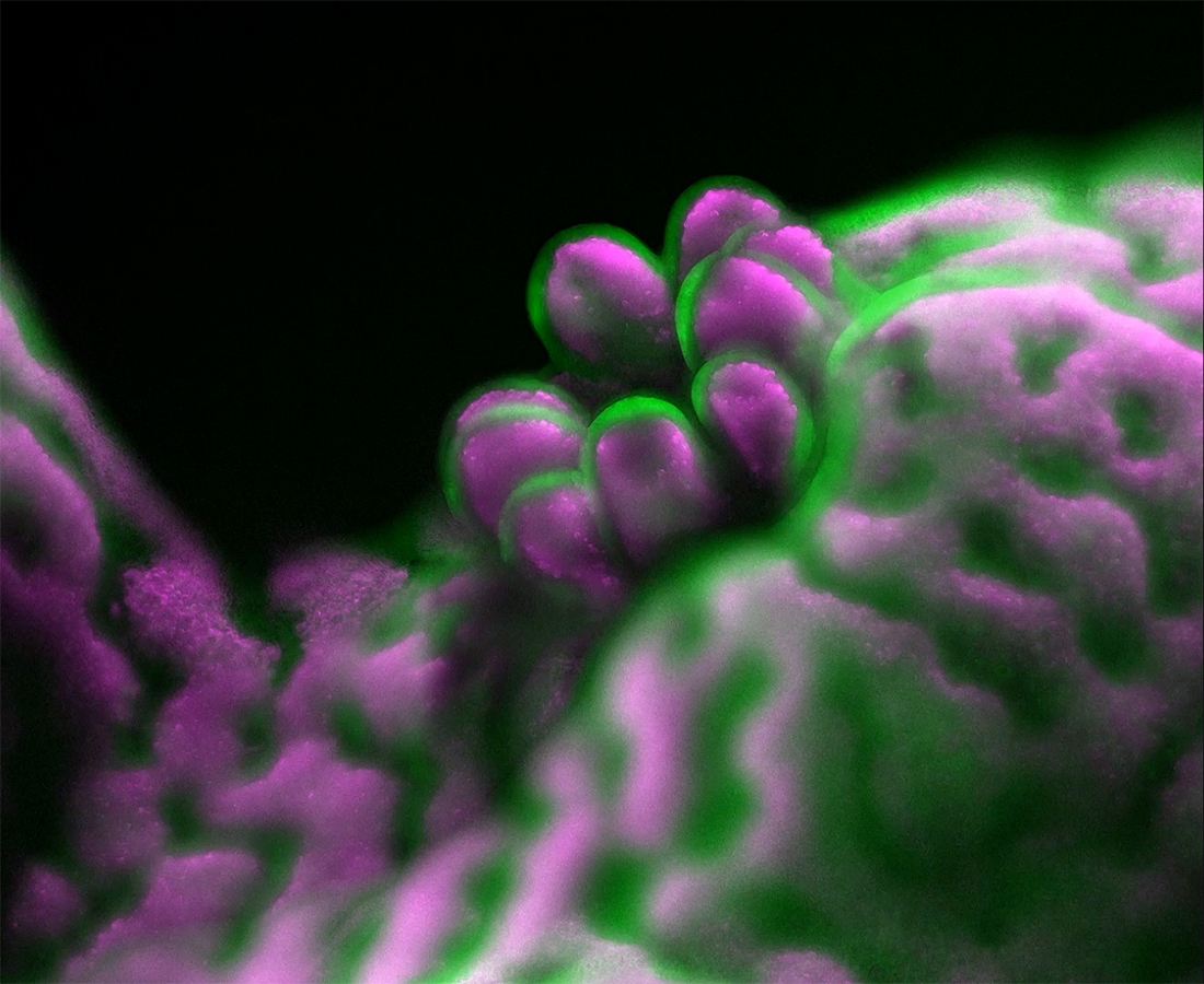
Nikon Instruments Inc. today revealed the winners of the ninth annual Nikon Small World in Motion Photomicrography Competition. Biologist and assistant professor/leturer Dr. Philippe Laissue captured the coveted top spot with his awe-inspiring video of a polyp emerging from a reef-building staghorn coral. Corals are extremely light sensitive, and capturing this video required Laissue to use a low-light technique and develop a custom microscope that could take movies of the corals without bothering the light-shy samples.
The video also captures the algae (colored in magenta) living inside the coral in a symbiotic relationship. Corals are made up of thousands of polyps and form coral reefs, which are an important part of marine ecosystems. The coral colonies Laissue is studying are essential reef-builders, but their sensitivity to bright light makes them very hard to film and study.
“Coral reefs are in alarming decline due to climate change, pollution and other human-made disturbances,” said Laissue. “I hope this video shows people the beauty of these organisms while raising awareness of their decline. We are working to better understand corals and their complex relationships with algae and other organisms. Hopefully we can contribute to finding the best ways to protect and conserve the coral reefs for future generations.”
“These amazing movies show us how much imaging technology has advanced over the years,” said Eric Flem, Communications Manager, Nikon Instruments, “It’s remarkable that we can bring stunning visuals like this one that highlight scientifically and socially relevant topics such as the decline of the reefs to the public.”
Laissue added, “Corals are utterly fascinating organisms - part animal, part plant, part stone. I’m grateful to be able to show the public a bit of their unseen world.”
Second place was awarded to Dr. Richard Kirby for his movie of Vampyrophrya, a type of parasite, emerging from their deceased host organism (a marine plankton). The video was captured using darkfield microscopy. Kirby says the most difficult part of capturing videos like this is transporting live samples from bodies of water to the laboratory for observation.
In third place is Tommy and Jesse Gunn for their video of a Stylonychia (microorganism) creating a water vortex using its cilia. The microscopic creature is creating this vortex in order to capture its next meal.
The 2019 judging panel included:
- Dr. Denisa Wagner, Edwin Cohn Professor of Pediatrics at Harvard Medical School and the head of the Wagner Lab at Boston Children’s Hospital.
- Dr. Rita Strack, Senior Editor at Nature Methods.
- Tom Hale, Staff Writer at IFLScience.
- Ben Guarino, Science Reporter at The Washington Post.
- Eric Clark (Moderator), Research Coordinator and Applications Developer at the National High Magnetic Field Laboratory at Florida State University
NIKON SMALL WORLD IN MOTION WINNERS
1st Place
Dr. Philippe P. Laissue
University of Essex
School of Life Sciences
Colchester, Essex, United Kingdom
Emerging Acropora muricata (staghorn coral) polyp (coral tissue in green; algae in magenta)
Custom-built Light Sheet Fluorescence Microscopy
10x (Objective Lens Magnification)
2nd Place
Dr. Richard R. Kirby
The Plankton Pundit
Plymouth, Devon, United Kingdom
Vampyrophrya (parasite) tomites swimming rapidly around within the body of the dead copepod host
Darkfield
1x (Objective Lens Magnification)
3rd Place
Tommy Gunn & Jesse Gunn
New York, New York, USA
Stylonychia (microorganism) creating a water vortex using its cilia
Darkfield
10x (Objective Lens Magnification)
4th Place
Dr. Hunter N. Hines
Harbor Branch Oceanographic Institute
Fort Pierce, Florida, USA
Two freshwater tardigrades feeding on another tardigrade
Differential Interference Contrast
10x (Objective Lens Magnification)
5th Place
Dr. Kate McDole & Dr. Philipp Keller
Howard Hughes Medical Institute
Janelia Research Campus
Ashburn, Virginia, USA
Developing mouse embryo, showing the progression of neural tube folding and closure
Light Sheet, Fluorescence
16x (Objective Lens Magnification)
HONORABLE MENTIONS
Thomas Drolsum
New Berlin, Wisconsin, USA
Iron filings in a magnetic field
Light Microscopy
10x (Objective Lens Magnification)
Caleb Foster
Caleb Foster Photography
Jericho, Vermont, USA
Reversed timelapse of a sublimating snowflake
Light Microscopy
4x (Objective Lens Magnification)
Dr. Jesse Gatlin, Abdullah Bashar Sami, Dr. John Oakey & Dr. April Kloxin
University of Wyoming
Department of Molecular Biology
Laramie, Wyoming, USA
Microtubule aster confined in a photo-patterned micro-container
Confocal
60x (Objective Lens Magnification)
Raul M. Gonzalez
Hiperfocal
Mexico City, Mexico
Hydroids
Darkfield
2.5x (Objective Lens Magnification)
Raul M. Gonzalez
Hiperfocal
Mexico City, Mexico
Caprellid (skeleton shrimp)
Darkfield
2.5x (Objective Lens Magnification)
Edwin Lee
Carrollton, Texas, USA
Radial canals emptying into a circular water-expelling vacuole in a protozoan
Phase Contrast
40x and 100x (Objective Lens Magnifications)
Dave R. Lewis
Clun, Shropshire, United Kingdom
Developing Rana temporaria (common frog) embryo (days 10 to 13)
Light Microscopy
4x (Objective Lens Magnification)
Dr. Kate McDole, Dr. Kristin Branson, Andrew Berger, Dr. Srinivas Turaga & Dr. Philipp Keller
Howard Hughes Medical Institute
Janelia Research Campus
Ashburn, Virginia, USA
Auto-detection of dividing cells in an entire developing mouse embryo
Light Sheet, Fluorescence
16x (Objective Lens Magnification)
Ani Michaud, Jiaye "Henry" He, Dr. Bill Bement, Dr. Jan Huisken & Dr. George von Dassow
University of Wisconsin-Madison
Laboratory of Cell and Molecular Biology
Madison, Wisconsin, USA
Traveling waves of protein activity in a single frog cell. The patterns form spontaneously and are involved in priming the cell for a future division.
Multiview Selective-plane Illumination Microscopy
10x (Objective Lens Magnification)
Dr. Andrew S. Moore & Dr. Erika Holzbaur
Howard Hughes Medical Institute
Janelia Research Campus, Janelia Advanced Imaging Center
Ashburn, Virginia, USA
Actin dynamics in a dividing cell
Lattice Light Sheet Microscopy
25x (Objective Lens Magnification)
Dr. Patrick Charles Nahirney
University of Victoria
Division of Medical Sciences
Victoria, Brititsh Columbia, Canada
Rare 'trinary' cell division (3 daughter cells) in L6 myoblast culture
Phase Contrast
10x (Objective Lens Magnification)
Wojtek Plonka
Krakow, Malopolskie, Poland
Formation of silver dendrites
Brightfield, Epi-illumination
3x (Objective Lens Magnification)
Caroline Pritchard
Lehigh University
Biological Sciences
Bethlehem, Pennsylvania, USA
Doryteuthis pealeii (longfin inshore squid) tentacle chromatophores (pigment cells)
Brightfield
4x (Objective Lens Magnification)
Dr. Shinji Shimode
Yokohama National University
Manazuru Marine Center
Manazuru-machi, Kanagawa, Japan
Marine planktonic larva of polychaeta worm
Rottermann Contrast
2x-6x (Objective Lens Magnifications)
Kar Yan Soh
The University of Auckland
Department of Molecular Medicine and Pathology
Auckland, New Zealand
Neutrophils (red) inside the hindbrain of a zebrafish larvae infected with green fluorescent bacteria
Confocal
20x (Objective Lens Magnification)
Johann Swanepoel
Justpixels
Randburg, Gauteng, South Africa
Stink bug hatching from egg
Reflected Light
10x (Objective Lens Magnification)
Dr. Sally Warring
American Museum of Natural History
Department of Genomics
New York, New York, USA
A ciliate from genus Pseudomicrothorax devours a cyanobacterial filament
Brightfield
20x and 40x (Objective Lens Magnifications)