2018 Photomicrography Competition
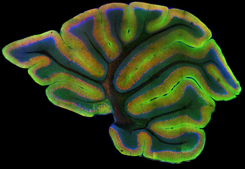
Cerebellum showing Purkinje cells (red), nuclei (blue) and tau (green)

Gabriel Luna Dr. Israel Hernandez, Dr. Kenneth S. Kosik

Affiliation
UC Santa Barbara
Neuroscience Research Institute
Santa Barbara, California, USA
Technique
Confocal
Magnification
40x (objective lens magnification)

Drosophila melanogaster (fruit fly) larval brains expressing photoconvertible EOS-tagged tubulin and showing neuronal connections

Dr. Wen Lu Dr. Vladimir I. Gelfand

Affiliation
Northwestern University
Feinberg School of Medicine
Department of Cell and Molecular Biology
Evanston, Illinois, USA
Technique
Fluorescence
Magnification
40x (objective lens magnification)

3D reconstruction of mouse testis tissue (green) and blood vessels (red)

Dr. Simon Frederik Merz Sebastian Korste, Sophie Henneberg

Affiliation
University Hospital Essen
Institute for Experimental Immunology and Imaging
Essen, Nordrhein-Westfalen, Germany
Technique
Light Sheet Fluorescence Microscopy and Tissue Clearing (Ethyl Cinnamate)
Magnification
2x (objective lens magnification)