2018 Photomicrography Competition
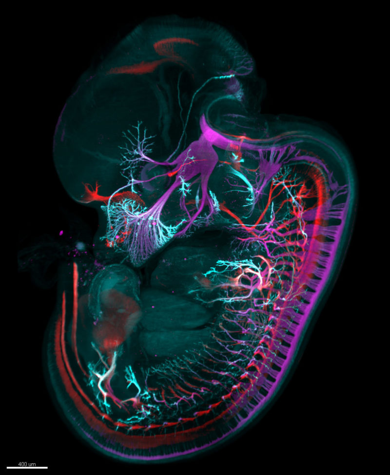
Image of Distinction
Mouse embryo (day 12.5) stained for motor (red) and sensory (magenta) nerves and nerve endings (cyan)
Dr. Gist F. Croft Lauren Pietila , Dr. Ali H. Brivanlou
- Affiliation
- The Rockefeller University
Laboratory of Stem Cell Biology and Molecular Embryology
New York City, New York, USA
- Technique
Light Sheet Microscopy and Tissue Clearing (iDISCO)
- Magnifaction
- 1.8x (objective lens magnification)