2018 Photomicrography Competition
5th Place
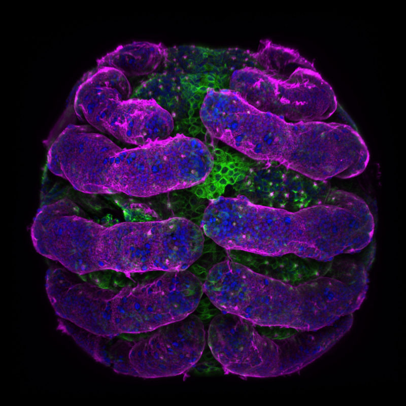
Parasteatoda tepidariorum (spider embryo) stained for embryo surface (pink), nuclei (blue) and microtubules (green)
Dr. Tessa Montague
- Affiliation
- Harvard University
Department of Molecular and Cellular Biology
Cambridge, Massachusetts, USA
- Technique
- Confocal
- Magnifaction
- 20x (objective lens magnification)