2025 Photomicrography Competition
10th Place
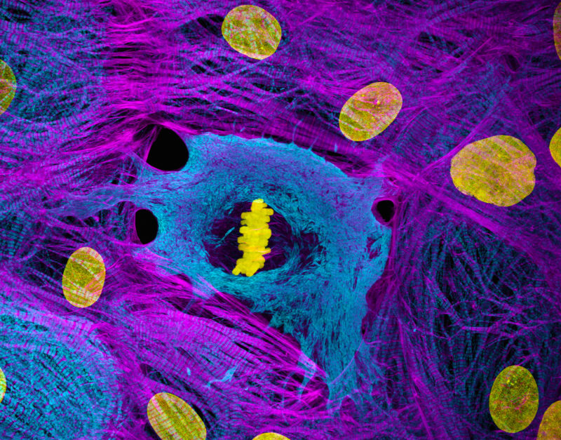
Heart muscle cells (iPSC-derived) showing condensed chromosomes in metaphase
Dr. Dylan T. Burnette Dr. James Hayes
- Affiliation
- Vanderbilt University
Department of Cell and Developmental Biology
Nashville, Tennessee, USA
- Technique
- Structured Illumination Microscopy (SIM)
- Magnifaction
- 60X (Objective Lens Magnification)
In Their Own Words
This picture shows human heart muscle cells that were grown from stem cells in the lab. Heart muscle cells rarely go through cell division. In the moment captured here, one cell is right in the middle of splitting into two. The bright yellow structure in the middle of the image are its chromosomes (bundles of DNA) lined up neatly in the center of the cell. This stage is called metaphase, and it’s the point where the cell double-checks that everything is correctly organized before it pulls the chromosomes apart and the mother cell completes the division into two new daughter cells.
What I love about this image is that it reveals something we rarely get to see heart muscle cells caught mid-division with their chromosomes perfectly aligned. It’s a reminder that even highly specialized cells that rarely divide can surprise us, and that the “invisible” processes inside our tissues are full of structure, drama, and beauty.