In this article, you’ll meet the members of the 2023 judging panel who reviewed thousands of microscopy submissions from across the globe to select this year's photo and video winners.
Meet the JudgesMeet the Judges
Meet the 2023 Judging Panel
Nikon Small World showcases brilliant imagery and artistic feats in science, and the unseen world around us. With every new year, the Nikon Small World judging panel plays an integral role in selecting the images and videos that best blend science and artistry. Nikon strives to select a panel that exemplifies a range of expertise – a team that can evaluate a photo or video based on technical prowess, scientific significance, aesthetic appeal, and power to convey a story to the viewer.
Ed Cara
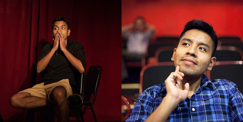
Ed Cara moonlighting as an improvisational comedian
As a Science Writer at Gizmodo, Ed Cara appreciates visual media that can tell a story. Whether it’s a spider’s fangs or pond creatures, these "fearsome” predators can offer some insight into the tiny domain that they claim as home.
Science and art have always been fundamental pillars in Cara’s life, leading him to pursue a bachelor’s degree in both theater and psychology at the University of Buffalo. Since graduating, Cara has channeled his passions into a career as a journalist with publications such as the Atlantic, Vice, Pacific Standard, and Undark Magazine.
For the past six years, Cara has painted a vivid portrait for his readers on topics ranging from public health to disease and weird animal science at Gizmodo. It felt only natural for Cara to bring his media expertise and love of all things science to this year’s Nikon Small World Competition.
“At the heart of it, both science and art are ways that we can try to understand the world around us,” said Cara. “It’s great when one can inform the other and spark the interest of the public so they can dig deeper and learn something new.”
When Cara is not writing his next big story or selecting winning microscopy images, he enjoys running, moonlighting as an improvisational comedian and volunteering. You can find his work here.
James Cutmore
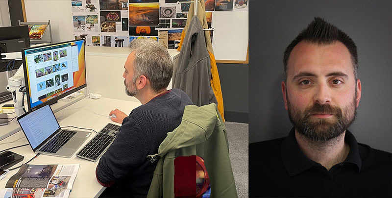
James Cutmore working on a story for BBC Science Focus Magazine
James Cutmore had always envisioned himself pursuing a career in physics or biology. But life had a different plan, and he decided to focus his energy on a bachelor’s degree in fine arts from the University of the West of England. He embarked on a picture editing role immediately after he graduated and for the last 19 years, he’s offered his unique background and innate creativity to BBC Science Focus Magazine.
Over the years, his role has diversified and allowed Cutmore to help grow BBC Science Focus Magazine’s brand by producing more online content and expanding into video production. “We are all about making science more accessible to people of all ages and levels of knowledge, and we are all very passionate about reaching as many people as we can,” said Cutmore. “I believe that science imagery is at the forefront of technology – it is the prime field where we see things in ever more detail, either under the microscope or in distant galaxies. It’s this constant change that makes every day different.”
His love of science imagery coupled with his historical knowledge of the industry made Cutmore an ideal candidate for the 2023 judging panel. “We humans are naturally curious, and we never stop learning,” said Cutmore. “Science can be dauntingly complicated when you don’t already have that knowledge, but through imagery and video we are able to communicate the wonders of the world we live in a way that people can understand. That can be the spark that ignites a life-long passion and can lead to a growing audience and more and more people getting involved in science!”
When reflecting on this year’s in-person judging at the Marine Biological Lab in Woods Hole, Massachusetts, Cutmore was delighted to serve as a judge. “I want to thank Nikon for giving me the opportunity to be involved with the Small World Competition. It has excelled at bringing science imagery to a wider audience through stunning images for nearly 50 years, and to play a part in that despite being a humble picture editor from a small town in the UK is a massive honor!”
You can find Cutmore’s work here.
Dr. Gary Laevsky
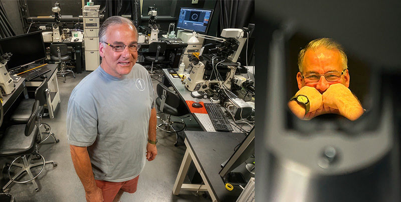
Dr. Gary Laevsky in his lab at Princeton University
Dr. Gary Laevsky always considered himself a “non-traditional student.” Before earning a Ph.D. in cell biology from the University of Connecticut in 2003, he spent the majority of his life working in restaurants and emergency medicine. After completing his post-doctoral research at The Scripps Research Institute in La Jolla, California, he called on the people skills he acquired working in service industries to shift into a client-facing career as a Product Manager for Olympus, a Senior Biosystems Applications Manager for Nikon, and an Imaging Applications Specialist for Andor Technology.
With a wealth of experience, Dr. Laevsky joined Princeton University in 2013 as Director of the Confocal Imaging Facility and received the title of Senior Professional Specialist, the highest non-faculty level position that can be achieved. “As the Director, I leverage my knowledge of the microscope industry to provide Princeton with access to state-of-the-art instruments, while teaching the newest generation of scientists how to use the available technology,” said Dr. Laevsky.
His unique career path and deep knowledge of all things imaging made Dr. Laevsky an indispensable member of the 2023 judging panel. “I think my expertise in the imaging world, in a multi-disciplinary environment, allows me to have a very well-rounded view of the capabilities the imaging community has to offer,” said Dr. Laevsky.
Upon reflecting on his experience as a judge, he enjoyed the camaraderie between his fellow panel members and contributing to such a noble competition. “These images allow the public to see, in a visually dramatic manner, what science is capable of achieving,” said Dr. Laevsky.
Among other achievements, Dr. Laevsky is a co-founder of the North Atlantic Microscopy Society (NAMS), Course Laboratory Director for the Analytical and Quantitative Light Microscopy (AQLM) course at the Marine Biological Laboratory (MBL), and co-founder of the Light-Sheet Fluorescence Microscopy (LSFM) Conference and Workshop, also at MBL.
Dr. Igor Siwanowicz
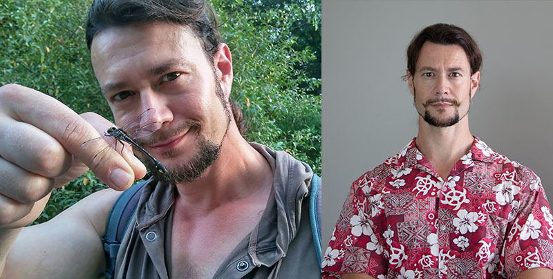
Dr. Igor Siwanowicz with a dragonfly
For Dr. Igor Siwanowicz, curiosity and an interest in biology came long before any thoughts of an actual career in the subject. Both of his parents were biologists, and he spent the better part of his childhood browsing through illustrated textbooks such as Ernst Haeckel’s “Art Forms in Nature” (long before he could read). His curiosity and desire to see the bigger picture led him to biochemistry and soon after, a new career path in neurobiology.
This transition was facilitated by his expertise in invertebrate morphology, which he developed through macro photography. A confocal microscope became his key tool of the trade, allowing for even more intimate insight into natural forms than a macro lens. For the past 10 years, Dr. Siwanowicz has been studying the anatomy and biomechanics of movement in invertebrates at the Janelia Research Campus of Howard Hughes Medical Institute in Ashburn, Virginia.
Along with his impressive background, Dr. Siwanowicz has placed a total of a total of 25 times in Nikon Small World and other scientific imaging competitions. “Microscopy can awaken viewers to the beauty of natural forms and the many facets of design that can be found in nature. It has the power to incite interest and curiosity and motivate some passionate investigations into the natural world.” said Dr. Siwanowicz.
Serving as a judge for this year’s competition was an unforgettable experience for Dr. Siwanowicz, “I’ve met and got to hang out with some truly interesting, genuine people that don’t take themselves too seriously and share my interest in microscopy – that always makes me happy. As a contributor to Nikon Small World for the past decade, I was in contact with some of them on many occasions. I finally got an opportunity to meet them in person. The lovely venue – Woods Hole is legendary at my workplace, and it was my first visit there – and the novelty of the whole situation made it a truly memorable experience.”
He went on to add, “The level of detail revealed by the microscope also often comes as a surprise – I had no idea that the appendages of a common brine shrimp (or a sea monkey) have such an intricate, almost fractal-like complexity, or that pollen grains of common asters look like a head of a morning star (a medieval mace).”
Dr. Clare Waterman
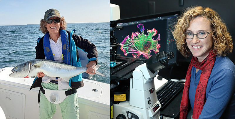
Dr. Clare Waterman on a recent fishing trip
Dr. Clare Waterman has had a great deal of experience when it comes to identifying visually compelling images for the Nikon Small World Competition. Having judged eight competitions, Dr. Waterman is known for her ability to understand difficult scientific techniques. “I’m always looking for technical innovation and whether it reveals something that you see every day but in a way that you have never seen before,” said Dr. Waterman. “Microscopy does that in general and sometimes it is really shocking to see how ordinary things, such as bug mouths or dog hair, look when magnified 1000 times.”
Regarded as a leading cell biologist in the scientific community, Dr. Waterman has more than 25 years of experience in imaging and microscopy. She graduated from Mount Holyoke College with a B.A. in biochemistry in 1989 and received an M.S. in exercise science in 1991 from the University of Massachusetts. “I had this idea that I would be a strength trainer for elite athletes. But when I saw electron microscope images of muscle for the first time, I thought that it was the most amazing thing I had ever seen in my life and decided that I wanted to do cell biology and microscopy.”
As her career took shape, Dr. Waterman received her Ph.D. in cell biology from the University of Pennsylvania in 1995. She spent 9 years as a professor in the Department of Cell Biology at the Scripps Research Institute in La Jolla, California. “I have always been interested in imaging living movement at the microscopic scale and that has been the focus of my career ever since.”
Each year, Dr. Waterman feels an immense honor in participating as a judge in the competition. “The thing I love about this contest is that it combines my two loves, art and science. It’s just a blast to see what kind of art people can produce in the medium that is my research tool.”
In her spare time, Dr. Waterman enjoys fishing in Woods Hole, Massachusetts.
To stay up to date with the "Meet the Judges" series and receive your daily dose of Nikon Small World, follow us on Instagram, Twitter, and Facebook. Be sure to follow Nikon Instruments for the latest updates on equipment and technology.