2018 Photomicrography Competition
11th Place
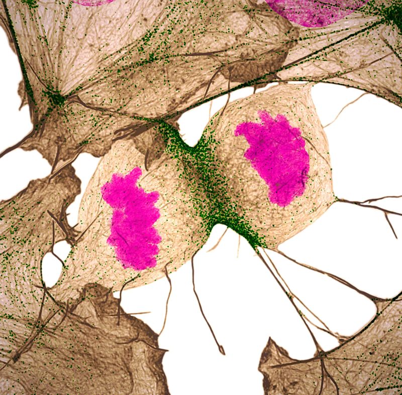
Human fibroblast undergoing cell division, showing actin (gray), myosin II (green) and DNA (magenta)
Nilay Taneja Dr. Dylan T. Burnette
- Affiliation
- Vanderbilt University
Department of Cell and Developmental Biology
Nashville, Tennessee, USA
- Technique
- Structured Illumination Microscopy
- Magnifaction
- 60x (objective lens magnification)