2020 Photomicrography Competition

Hebe plant anther with pollen

Dr. Robert Markus Zsuzsa Markus

Affiliation
University of Nottingham
School of Life Sciences, Super Resolution Microscopy
Nottingham, Nottinghamshire, United Kingdom
Technique
Confocal
Magnification
10X (Objective Lens Magnification)

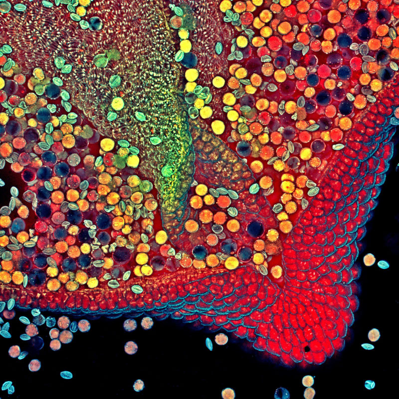
Dr. Robert Markus, a super-resolution and confocal microscopy senior imaging technician from the University of Nottingham, took this image of a Hebe plant anther with pollen in order to create awareness of the beauty of the natural world around us and to help showcase the performance of the latest microscope technology. It was captured using the highest numerical aperture lens for low magnification and confocal lasers set to mimic how the subject would look under UV light. It shows in very high detail anther pollen and the cell structures within the plant.

Bogong moth

Ahmad Fauzan

Affiliation
Saipem
Jakarta, Indonesia
Technique
Image Stacking
Magnification
5X (Objective Lens Magnification)

Mr. Ahmad Fauzan, an engineer by trade and microscopy hobbyist, created this image of a bogong moth with the goal of illustrating the intricate detail in this somewhat rare insect. This moth breed can take one of two forms, migratory and non-migratory, which are differentiated by the look of their hind wings. In order to capture the detail of the head, mouth and proboscis (shown), image stacking software was applied to 64 individual photographs comprising this winning picture.

Microtubules (orange) inside a BPAE cell. Nucleus is shown in cyan.

Jason Kirk

Affiliation
Baylor College of Medicine
Optical Imaging & Vital Microscopy Core
Houston, Texas, USA
Technique
Confocal
Magnification
63X (Objective Lens Magnification)

Jason Kirk, a professional microscopist and director of the Optical Imaging and Vital Microscopy Core facility at Baylor College of Medicine, created this image using a confocal microscope combined with a variation of image scanning microscopy. It was taken primarily to benchmark the performance of the microscope systems within the facility and beautifully shows the radial arrangement of microtubules in bovine pulmonary artery endothelial cells.