Jason Kirk, a professional microscopist and director of the Optical Imaging and Vital Microscopy Core facility at Baylor College of Medicine, created this image using a confocal microscope combined with a variation of image scanning microscopy. It was taken primarily to benchmark the performance of the microscope systems within the facility and beautifully shows the radial arrangement of microtubules in bovine pulmonary artery endothelial cells.
2020 Photomicrography Competition
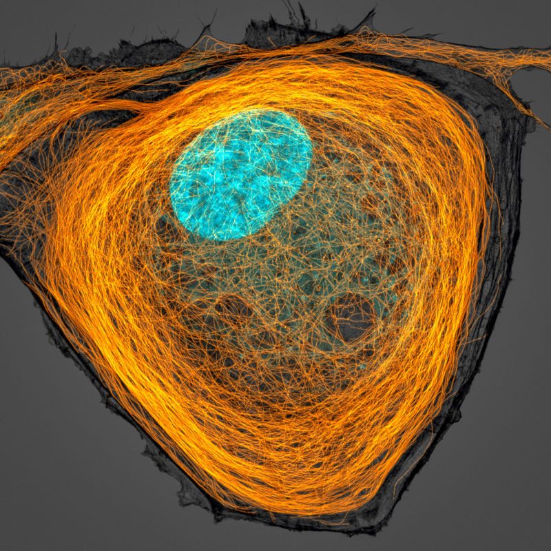
7th Place
Microtubules (orange) inside a BPAE cell. Nucleus is shown in cyan.
Jason Kirk
- Affiliation
- Baylor College of Medicine
Optical Imaging & Vital Microscopy Core
Houston, Texas, USA
- Technique
- Confocal
- Magnifaction
- 63X (Objective Lens Magnification)