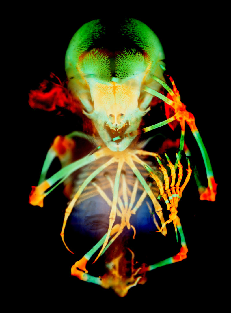
This image taken by Dorit Hockman of a skeleton of a short tailed fruit bat embryo beautifully illustrates the details of the elongated hand bones that form the scaffold of the bat wing. This x-ray-like image took months to prepare, with the sample preparation being done by Dr. Vanessa Chong-Morrison at the Marine Biological Laboratory in Woods Hole, Massachusetts. The sample took months of waiting for the specimen to be clear enough to reveal the bones, upon which the task of capturing this image began. It was a composite of images taken at high magnification and stitched together to the final image on display.
2020 Photomicrography Competition
20th Place
Skeleton preparation of a short-tailed fruit bat embryo (Carollia perspicillata)
Dr. Dorit Hockman Dr. Vanessa Chong-Morrison
- Affiliation
- University of Cape Town
Cape Town, Western Cape, South Africa
- Technique
- Brightfield
- Magnifaction
- 1X (Objective Lens Magnification)