Dr. Grigorii Timin on the Hidden Beauty and Complexity of the Gecko
Welcome to Masters of Microscopy: The People Behind the Lens, where we showcase and celebrate the individuals who are the heart of the Nikon Small World competitions. They are scientists, artists, researchers, educators, and everyday curious individuals who uncover the fascinating microscopic world around us.
Dr. Grigorii Timin’s passion for microscopy started at the age of 15 when he was gifted a microscope and explored the creatures living in a pond close to his summer home. Timin spent his vacations observing protists, worms and diatom algae and learning about the world around him.
After completing a master’s degree in Biophysics at the Saint-Petersburg Polytechnic University, his sense of discovery and deep-seated curiosity led him to the Ph.D. program at the University of Geneva. Under the supervision of Dr. Michel Milinkovitch, Timin focused his time on the genetics and mechanics of morphogenesis, the process by which tissues and organs acquire their shapes and physical forms. In particular, he studied the development and evolution of skin appendages in reptiles.
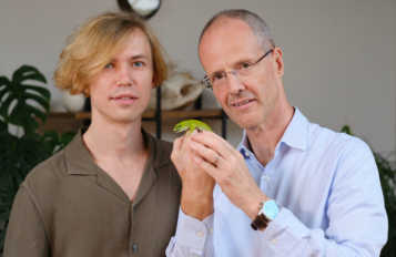
Dr. Grigorii Timin and Dr. Michel Milinkovitch with an adult Madagascar giant day gecko (Phelsuma grandis)
To advance the study of morphogenesis of scales in snakes, Timin devised a new method to image the 3D network architecture of collagen and demonstrate how a network is organized.
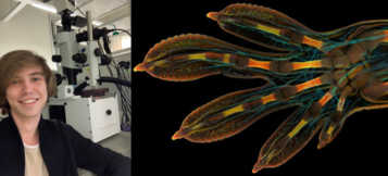
“The image that I submitted to the Nikon Small World competition was one of the samples that I used to prove this new method,” said Timin. “I was able to visualize the collagenous structures in a whole developing organ in precise detail and on a large scale.”
Alongside his new method, Timin used image-stitching to merge hundreds of images together to create the final image of his gecko. Preparing the sample was an added challenge. Timin performed whole-mount fluorescent staining and tissue clearing to capture the entire embryonic hand with a confocal microscope.
"This embryonic hand is about 3 mm (0.12 in) in length, which is a huge sample for high-resolution microscopy," said Timin. "The scan consists of 300 tiles, each containing about 250 optical sections, resulting in more than two days of acquisition and approximately 200 GB of data."
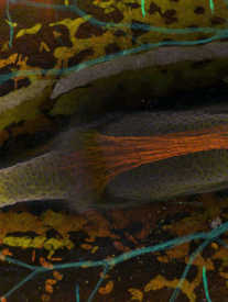
A magnified region of Timin’s winning image
The winning image highlights the nerves in a cyan color and the bones, tendons, ligaments, skin and blood cells in a range of warmer colors. "This particular image is beautiful and informative, as an overview and also when you magnify it in a certain region, shedding light on how the structures are organized on a cellular level," said Timin.
Reflecting back on this year’s competition, Timin shared, “The Nikon Small World competition is a great opportunity to share how impressive nature is on a microscopic level, not only within a scientific community but also with the general public. Few people have a chance to look at things under a microscope, and for the most part, they are pretty beautiful.”
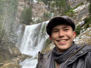
When Timin is not exploring new worlds and sharing his images with the public, he enjoys the classical and alternative music scenes with friends in Geneva and discovers picturesque views of Switzerland nature.
Timin enjoying Switzerland nature
To stay up to date with the "Masters of Microscopy" series and receive your daily dose of Nikon Small World, follow us on Instagram, Twitter, and Facebook. Be sure to follow Nikon Instruments for the latest updates on equipment and technology.