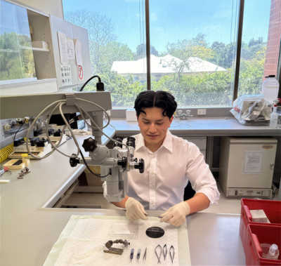
For Hassanain Qambari, his passion for microscopy developed as a student at the University of Western Australia (UWA) and The Lions Eye Institute under the mentorship of Professor Paula Yu, Professor Dao-Yi Yu and Professor Chandrakumar Balaratnasingam. Qambari earned a bachelor's and master's degree in biomedical science specializing in neuroscience at UWA and is on track to earn his Ph.D. Currently, Qambari is investigating the early changes in the endothelium and its reversal in early diabetic retinopathy.
Masters of MicroscopyMasters of Microscopy
Hassanain Qambari on Revealing the Intricacies of Diabetic Retinopathy
Welcome to Masters of Microscopy: The People Behind the Lens, where we showcase and celebrate the individuals who are the heart of the Nikon Small World competitions. They are scientists, artists, researchers, educators, and everyday curious individuals who uncover the fascinating microscopic world around us.
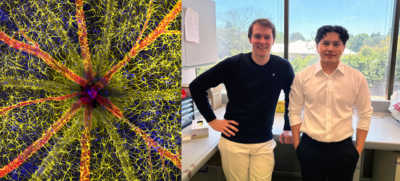
Qambari’s winning image; taken with the assistance of Jayden Dickson
Diabetic retinopathy occurs when high blood sugar damages the blood vessels in the tissue at the back of the eye, known as the retina. The damaged blood vessels can swell and leak, which can cause blurry vision or total loss of eyesight. Since 2021, Qambari has devoted his time and research to the early detection and reversal of the disease.
Assisted by Jayden Dickson, Qambari won this year’s Nikon Small World Photomicrography competition for his vivid image of a rodent optic nerve head showing astrocytes (yellow), contractile proteins (red), and retinal vasculature (green). The colorful image provides an important contribution to the study and reversal of diabetic retinopathy.
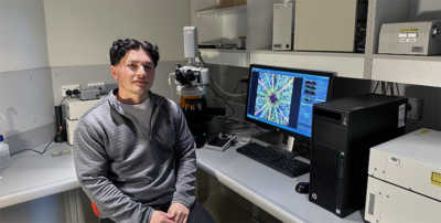
Qambari beside his winning image and a confocal microscope system
“Current diagnostic criteria and treatment regimens for diabetic retinopathy are limited to the late-stage appearance of the disease, with irreversible damage to retinal microvasculature and function,” said Qambari. “The visual system is a complex and highly specialized organ, with even relatively minor perturbations to the retinal circulation able to cause devastating vision loss. I entered the competition as a way to showcase the complexity of retinal microcirculation.”
It was no easy task to capture his winning image and at some points, a technically demanding challenge. After locating the fine vessels near 110 microns in diameter, Qambari established a protocol for labeling different cell types. “Over the past 20 years, our research group has refined the technique of isolated ocular perfusion labeling for fine vessels in the eye,” said Qambari. “While the ophthalmic artery in the rodent model presented a challenge, we were able to overcome it with persistence and patience.”
For participants like Qambari, the competition is an opportunity to share new perspectives with a wider audience. “The Nikon Small World competition is great, as it showcases amazing work across many disciplines from around the world,” said Qambari. “All the images presented in the competition represent the beauty and artistic side of science which may otherwise get overlooked. Such a competition not only celebrates the participant's hard work and passion but may also draw and inspire young scientists to pursue a career in STEM. It certainly inspired me.”
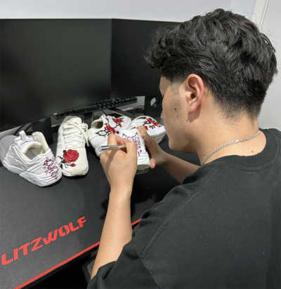
Qambari customizing sneakers
Outside the lab, Qambari enjoys sports such as soccer and badminton, customizing sneakers and building online games using the programming language of Python.
To stay up to date with the "Masters of Microscopy" series and receive your daily dose of Nikon Small World, follow us on Instagram, Twitter, and Facebook. Be sure to follow Nikon Instruments for the latest updates on equipment and technology.