When Small Worlds Collide: Collaboration Behind the Microscope with Liz Haynes and Henry He
Welcome to Masters of Microscopy: The People Behind the Lens, where we showcase and celebrate the individuals who are the heart of the Nikon Small World competitions. They are scientists, artists, researchers, educators and everyday curious individuals who uncover the fascinating microscopic world around us.
To film the developing embryo, Dr. Haynes and Mr. He chose to let the zebrafish embryo grow in water, where it develops naturally inside their home-built microscope. This is a much more challenging approach as the specimen could easily move out of the field of view. The conventional technique of mounting the zebrafish in a block of gel however, restricts the growth of the embryo, which can impact the development of the neurons and result in a less accurate study.
Since they began to work together at the Morgridge Institute for Research, the pair’s collaboration has moved beyond the laboratory. Combining Haynes’s love for the outdoors with Mr. He’s passion for photography, the two have taken up bird watching and hiking in their spare time. Their most recent outing was to capture the migration of Sandhill cranes as they return to the upper Midwest. “We’re not just collaborators, we really are friends,” said Haynes.
A photo taken by Mr. He on a bird-watching expedition with Dr. Haynes.
A Partnership in the Making
Haynes’s fascination with biology began at a young age. “There was never really any question for me at any point in time that I wanted to be a scientist,” she recalled, “I kept lizards at my house as a kid, and did all sorts of little experiments. I was this nerdy 14-year-old kid who was shooting my hand up in microbiology class because I wanted to answer everything.”
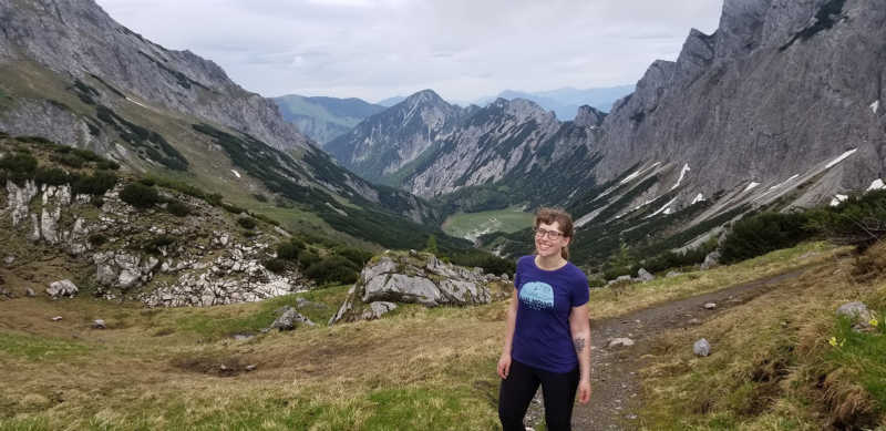
A partnership that became a friendship. Photo taken by Mr. He on a hiking trip with Dr. Haynes.
Haynes’s undergraduate and graduate education fueled this passion. “I started undergraduate research and everything clicked into place. I meandered into a lot of different realms in science, but found that I really loved what I could study visually.”
For He, microscopy wasn’t always top of mind. In fact, he began his scientific career as a mathematician and astrophysicist. While he was gifted with numbers and had a passion for research, it was the immediacy of biology that attracted him to the microscope: “It dawned on me that I preferred the pace of biological research. Astrophysics research cycles tend to last much longer, and I liked working on something where I could get a result back right away, which led me to focus on the biological sciences.”
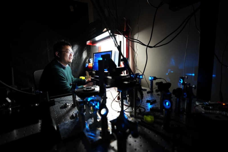
Mr. He conducting his biological research
He still wanted to do quantitative work; and wanted to focus on the technical process of biological research. “When I started searching for a Ph.D. program, I made the decision that I wanted to be a method developer. I want to develop the instrumentation, develop the protocol, and then optimize it.”
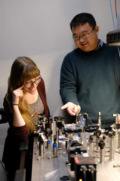
Far from putting them into separate scientific worlds, their different backgrounds and areas of study make Haynes and He the perfect research pair. At the time, Haynes’s neurobiology lab studied the development of the zebrafish nervous system, but ran into a slew of technical questions regarding microscopy and image optimization. “Henry's lab came in and we were able to push into large-scale imaging of the entire fish and capture live videos with light sheet technology,” said Haynes.
Collaborating with Haynes’s lab has allowed He to focus on what truly interests him: the imaging process. “Before moving to the University of Wisconsin, our lab was a hybrid: 60% of the lab were biologists and 40% were developers who built and optimized the instruments. Now that we’ve started working with Liz’s laboratory, our biological question is completely outsourced,” said He. “Now, our lab is focused more on instrument design and data analysis-- we’re probably at 20% biologists and 80% developers.”
This symbiotic relationship has provided new opportunities for Haynes and Henry to take stunning images, like those showcased in their award-winning Small World in Motion video. “I think the great part of our collaboration is that Henry and I have worked closely together from the outset,” said Haynes. “We designed our plan together and we set the parameters together.”
“I think it's a really great example of two people coming from two separate fields but both speaking the same language and working together to achieve something really great,” remarked Henry.
To stay up to date with the "Masters of Microscopy" series and receive your daily dose of Nikon Small World, follow us on Instagram, Twitter, and Facebook. Be sure to follow Nikon Instruments for the latest updates on equipment and technology.