Before Nancy Kedersha was an Instructor of Medicine at Harvard’s Brigham and Women’s Hospital, before she was an eminent scholar on stress granules and ribosomal proteins, and before she was a four-time Nikon Small World finalist and judge, she was a grade schooler with a passion for two subjects that she thought could never coincide: science and art.
Masters of MicroscopyMasters of Microscopy
Dr. Nancy Kedersha on the Art and Science of Sample Preparation
Welcome to Masters of Microscopy: The People Behind the Lens, where we showcase and celebrate the individuals who are the heart of the Nikon Small World competitions. They are scientists, artists, researchers, educators and everyday curious individuals who uncover the fascinating microscopic world around us.
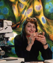
Dr. Kedersha with her scope
“I had a very pointed interest in science, but I also quite liked art. At the time, it never occurred to me that there was some way to do both,” recalls Kedersha. “It was always a question of am I going to do this? Or am I going to do that?”
Professionally, Dr. Kedersha opted for science, obtaining her Ph.D. from Rutgers University and pursuing a post-doctorate in the laboratory of Dr. Leonard Rome at UCLA. She remembers the startling beauty of observing an immuno-stained sample for the first time: “I was staining for prolyl hydroxylase and I looked and saw these expansive glowing galaxies, all contained in tiny, little cells. I thought: ‘wow, I really, really like this.”
As she continued her research into cancer-specific antibodies for immunotherapy (a process that involves using toxin-modified antibodies to target cancer cells) Nancy honed her expertise in imaging through extensive work with the processes of photobleaching, fluorescent tagging, using electron and light microscopy. Her prowess in immunostaining grew to the point where colleagues were constantly sending her images and preparations to evaluate.
“Immunostaining is a complex and delicate process,” says Kedersha. “You can do it right and still get the wrong answer. It’s like peeling an onion; you have to treat the preparation with just the right combination of permeabilization agents and fixation agents to let the antibodies get in and show you what’s there.”
The process is variable. It isn’t so much about memorizing a formula as it is listening to experience and learning from trial-and-error. “You really need to know your tools,” says Kedersha. “You have to understand how your tools work before you can figure out what the data is trying to tell you. You need to know, this fixation agent does this, but not when mixed with this permeation agent. You need to know the ins-and-outs of each piece to understand the result of the whole. Honestly, I don’t consider myself so much as a Master of Microscopy as I do a Master of Sample Preparation.”
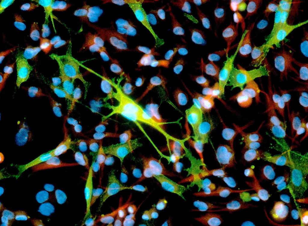
This micrograph of melanoma cancer cells won Nancy 2nd place in 1991
Understanding your tools and mastering the art and science of sample preparation can yield stunning results, such as Kedersha’s image of rat brain cells that won 3rd place in the 1989 Photomicrography competition, or her triple exposure of a melanoma and astrocytoma cell culture which took 2nd place in the 1991 competition. Kedersha has placed in the Nikon Small World competition a total of 10 times since she first began entering the competition in 1989.
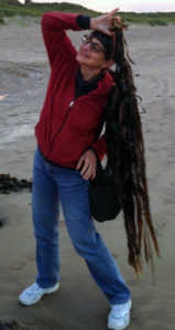
Now semi-retired, Kedersha loves to travel. Here she is enjoying the beach in Northern Ireland.
However, photomicrography is not the only discipline in which Nancy uses her tools to yield beautiful results. Kedersha refers to herself as a “rogue ecologist” as she tends to her garden at her home in Southern Maine. She utilizes her knowledge of biological processes to maintain her outdoor spaces in all-natural ways. Whether it’s bringing in a herd of goats to eat the invasive bittersweet vines off of her trees or introducing native fish to eat the leeches in her pond, Kedersha knows how to get the most of the biological tools at her disposal.
According to Kedersha, it was microscopy competitions like Nikon Small World that showed her that her two passions, science and art, really could coexist. “Doing microscopy made me realize that I can do art and science at the same time. I can implement the rigor of science and still have an aesthetic dimension that can inspire people. So, am I going to do this? Or am I going to do that? I did both.”
To stay up to date with the "Masters of Microscopy" series and receive your daily dose of Nikon Small World, follow us on Instagram, Twitter, and Facebook. Be sure to follow Nikon Instruments for the latest updates on equipment and technology.