Philippe Laissue on Capturing Sensitive Creatures and Working with Living Things
Welcome to Masters of Microscopy: The People Behind the Lens, where we showcase and celebrate the individuals who are the heart of the Nikon Small World competitions. They are scientists, artists, researchers, educators and everyday curious individuals who uncover the fascinating microscopic world around us.
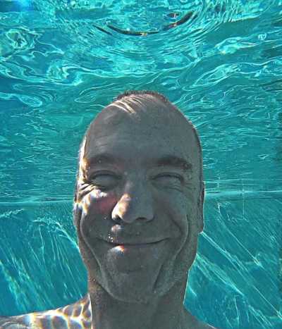
From snorkeling to microscopy, there’s nothing Dr. Philippe Laissue loves more than diving into a world beyond our view.
As a lecturer at the University of Essex, that’s something he gets to do with his students every day. Whenever possible, however, he also loves to share these worlds beyond the classroom. To do that, Laissue has dedicated much of his work to the art and science of bioimaging, and has even developed his own microscope.
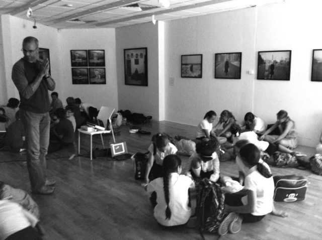
Laissue teaches a class of science students
“While we use standard laboratory techniques for our research, I have to admit that the single most gratifying thing is getting an image or a movie as a result of an experiment,” Laissue says.
The result of these efforts has led to some incredible imagery. Laissue has placed twice in recent years for these images in Nikon’s Small World Competition, and took the first prize this year for his video of a polyp of a reef-building staghorn coral. The video shows in unprecedented detail the dynamics of a fluorescent coral polyp and the algae living inside its tissue, over a period of eight and a half (or ‘nearly nine’) hours.
1st Place, 2019 Small World in Motion Competition
“Corals are utterly fascinating organisms - part animal, part plant, part stone. It is easy to see why people could not decide for millennia what they were. In my movie, a polyp emerges like a blossoming flower,” Laissue says.
Laissue’s approach to photographing coral is unique. Because the light is low, the polyp emerges fully. “The small coral colonies I image are highly photosensitive, they do not like bright light. So it was necessary to use a low-light imaging technique. I had to build a custom light-sheet microscope that could take movies of these large, light-shy samples,” he says.
When Laissue first wanted to look at coral, he attempted to use a confocal spot scanner, but was only able to get one image before the coral began reacting to the bright light. He then switched to widefield illumination, which gives less intense light, but he wasn’t able to get that nice optical sectioning that he had with the confocal.
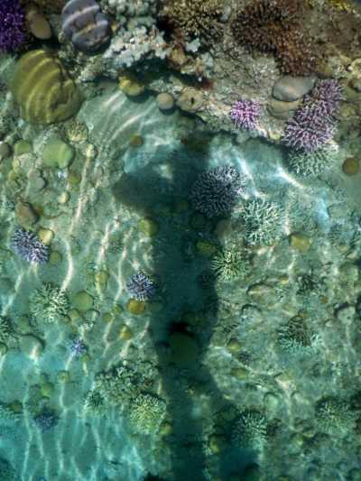
Laissue taking some macro-photography photos of a reef
“So I combined the best of both worlds with light-sheet microscopy, where you get the optical sectioning together with the very gentle illumination,” says Laissue. “However, I needed to make one that could take small coral colonies, which by microscopy standards are huge samples.”
For anyone who may just be getting into bioimaging, Laissue recommends starting with macrophotography. When you’re ready to move to microscopy, Laissue emphasizes learning about light and its effect on your sample. “If you're looking at something living, I think people often don't worry, and need to be more aware of the risk of damaging the sample with the light that they are using,” he says.
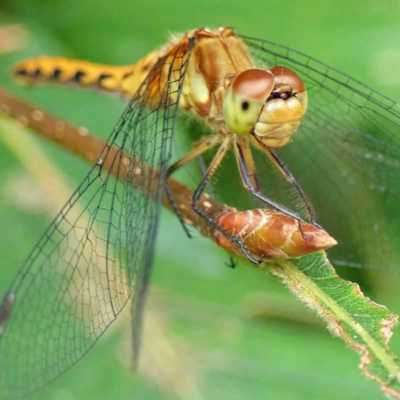
Laissue doesn’t just love to study corals, his interests also expand to insects like this dragonfly (Orange Meadowhawk (Sympetrum))
Today, Laissue’s main research interest continues to be about understanding that risk and in finding innovative ways to detect phototoxicity – and then avoid it, or at least minimize it. He is also interested in finding out why and how the symbiosis between the coral and its algae breaks down, and in raising awareness of coral decline due to climate change, pollution, and other man-made disturbances.
And of course, he hopes to continue sharing the small worlds he discovers with new audiences. “As researchers, our main focus of microscopy is to extract numbers from the structures and events we image and to analyze them scientifically. But it is also important to remind ourselves of their beauty - and share it with the world This is a wonderful thing to do, especially with kids, who are naturally very curious. We humans are such visual creatures,” he adds. “We absorb over 90% of information through our eyes, so being able to communicate science and research visually has in my view a big impact.”
To stay up to date with the "Masters of Microscopy" series and receive your daily dose of Nikon Small World, follow us on Instagram, Twitter, and Facebook. Be sure to follow Nikon Instruments for the latest updates on equipment and technology.