Welcome to Masters of Microscopy: The People Behind the Lens, where we showcase and celebrate the individuals at the heart of the Nikon Small World competitions. They are scientists, artists, researchers, educators, and everyday curious individuals who uncover the fascinating microscopic world around us.
Dr. Ning Xu is a researcher in optical engineering who develops advanced imaging systems that help scientists observe cellular structures in motion. Working at the intersection of optics, biology, and technology, he focuses on building microscopes that capture tiny structures moving at exceptionally high speeds. It is this work that earned Xu first place in the 2026 Nikon Small World in Motion competition.
His winning video displays the movement of cilia, tiny hair-like structures found throughout the body's airways, in a patient-derived sample from a child with primary ciliary dyskinesia (PCD), a rare genetic disorder that affects how cilia move. PCD is revealed by the movement of cilia rather than by their appearance alone, so accurate diagnosis relies on dynamic imaging with highly specialized systems, such as the one impressively developed by Xu and his collaborators.
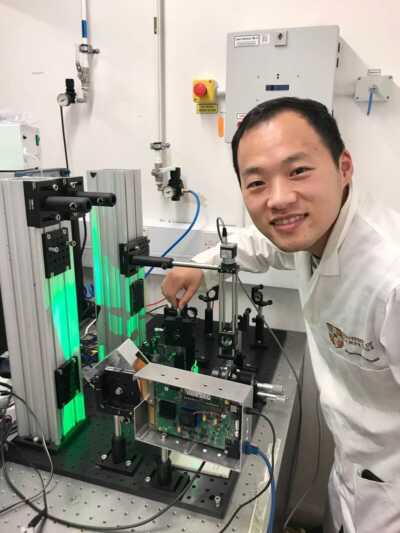
The second-generation diffractive super-resolution microscope system, 2021
Xu's path to microscopy began more than a decade ago, with a much simpler instrument. As an undergraduate, he participated in a competition that challenged students to build a microscope with a single lens. That microscope was rudimentary, and though he won fifth place, he says the images he captured changed his perspective.
"When I saw the first image from an instrument I had built with my own hands,” he added, “I only thought, ‘I could create another that makes things even clearer.’”
That curiosity eventually led to a career in optical engineering. Xu went on to develop increasingly sophisticated imaging systems, collaborating with researchers at Tsinghua University, the University of Cambridge, Beijing Children's Hospital, and the California Institute of Technology along the way. Today, as a research fellow at the National University of Singapore, he builds microscopes that combine advanced optical techniques with artificial intelligence to capture life as it actually happens: in motion.
“For over 50 years, Nikon Small World has repeated one simple message: science is beautiful,” Xu said. “And Small World in Motion has a second message that I'm even more passionate about: life is not a still picture. Life moves.”
Cilia are a good example of that movement. Millions of microscopic structures line airways, where they beat together in coordinated waves to move mucus and debris out of the lungs. In people with PCD, the cilia can move abnormally, making it difficult for the airways to clear themselves and contributing to recurrent respiratory infections. Capturing and studying that movement gives researchers another way to understand what is happening in the cells, and to diagnose it earlier and more accurately.
Recording the movement of cilia at the level Xu wanted to see posed a significant imaging challenge. Individual cilia are extremely small, and their movement is rapid. Plus, the samples need to remain alive and behave naturally during imaging. Many advanced microscopy techniques rely on fluorescent labels or intense illumination to make structures easier to see, but those approaches can be problematic in this situation, when the goal is to observe living tissue without disturbing it.
“Everyone wants a bright, clean image, but strong light damages living cells and changes their behavior,” he advised. “Capturing cells’ real, natural behavior is better and more important than producing a perfect video of dying cells.”
Xu and his collaborators instead focused on controlling the light itself. Their imaging system uses multiple light waves and a digital micromirror device to precisely shape and control illumination, allowing them to capture the rapid movement of cilia at high resolution while minimizing the light reaching the sample. A technical challenge, which Xu describes simply: “Fast, gentle, and sharp. The whole system must do all three at once.”
The system used to capture his winning video, recorded in December 2025, was the fifth version of the technology, following several years of development that began in 2021. Earlier versions of the system were developed with collaborators including Professor Sarah Bohndiek at the University of Cambridge and Dr. Calum Williams, now at the University of Exeter, whose work in biomedical optics helped shape the approach.
Xu and his collaborators knew they had accomplished something big, but Xu had no idea it would result in a first-place win.
“I grew up with Nikon Small World images. You see them in textbooks, in exhibitions, and all over the internet,” he said. “So, joining this tradition feels a little unreal, especially since this was my first time entering the competition. I never thought I could win.”
The winning video also represents something Xu finds important about scientific communication. Much of his work is ultimately presented through scientific papers, which can make it difficult for people outside the field to understand what researchers are seeing or why it matters. In this case, the movement of cilia offers a glimpse into a biological process occurring inside the human body that would otherwise remain completely invisible.
“When people watch cilia beating, they immediately understand that something beautiful and important is happening inside us,” Xu said. “I think this competition turns microscopy into a shared language.”
Xu's work has been shaped by several mentors throughout his career. He credits his Ph.D. advisor, the late Dr. Qiaofeng Tan, with teaching him how to design optics, shape light, and build optical instruments. He also cites Nobel laureate Dr. Eric Betzig as an important influence, particularly Betzig's persistence in finding new ways to see biological structures. His current supervisor, Dr. Changhuei Yang, believes in doing research that industry and clinicians can actually use, an approach that shaped Xu's decision to join his group, and one he now applies to exploring how imaging technology can be deployed in clinical pathology.
Winning Nikon Small World's 2026 Small World in Motion competition came at a transitional moment for Xu. He received the news while moving from Beijing to Singapore, leaving behind his position as a lecturer at Tsinghua University to join the National University of Singapore as a research fellow, making the recognition an unexpected highlight amid a major career change.
“It’s one of the proudest professional moments of my life,” he said.
Outside the lab, Xu enjoys cycling, hiking, and swimming, particularly when he is exploring a new place. “After long days in a dark imaging room, I need some sunlight!” he joked.
Hiking in the Tianshan Mountains in 2025
Jokes aside, microscopy continues to occupy much of his attention, and there is still plenty he wants to see.
“I need to study more and find more life in motion. I want to pursue that for the rest of my life,” Xu said.
While Xu has spent his career building increasingly sophisticated ways to see the microscopic world, he is still motivated by the same curiosity that led him to build his first microscope as a student. There is always another structure to resolve, another movement to capture, and another question that might become visible with the right combination of light and optics.
His advice to other researchers considering entering the competition is straightforward: “Take care of the sample, use as little light as possible when working with living cells, and most importantly, keep recording. The best moment usually appears right after you decide to stop.”