Nikon Small World Honors 51st Annual Photomicrography Competition
Posted on October 15, 2025
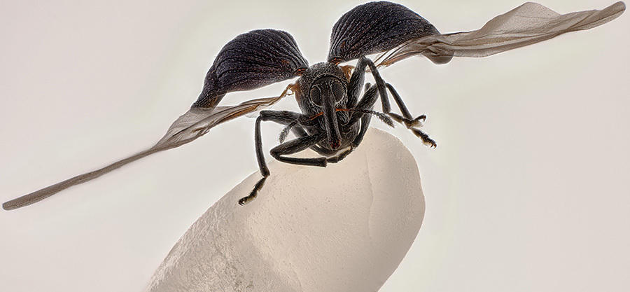
Nikon Instruments Inc. today announced the winners of the 51st annual Nikon Small World Photomicrography Competition, celebrating over five decades of excellence in microscopy and digital imaging. The first-place prize was awarded to China’s Zhang You for his striking image of a rice weevil mounted on a grain of rice. The image captures the insect with its wings fully extended, frozen in a moment that provides insight into the structure and behavior of a familiar yet often overlooked agricultural pest.
A member of the Entomological Society of China and the Entomological Society of Yunnan Province, You’s winning work is a product of the years he has spent focused on ecological and insect science photography, as well as teaching others about entomology. “It pays to dive deep into entomology: understanding insects’ behaviors and mastering lighting,” You said. “A standout work blends artistry with scientific rigor, capturing the very essence, energy, and spirit of these creatures.”
The choice of scale in the image emphasizes the insect’s actual size while contextualizing its ecological role as a pest known for attacking seeds of several crops. Using a medium-format camera paired with a 5x microscope objective, You captured over 100 images for focus stacking, carefully cleaning, lighting, and post-processing the specimen over the course of two weeks.
The subject itself was a rare and fortunate find. “I had observed rice weevils in grains before, but never one with its wings spread,” You explained. “This one was naturally preserved on a windowsill, perhaps in a final attempt to escape. Its tiny size makes manually preparing spread-wing specimens extremely difficult, so encountering it was both serendipitous and inspiring.” Insects, from pollinators to pests, play vital roles in ecosystems and economies alike, and You’s work encourages audiences to recognize the complexity hidden among these communities.
In addition to winning first place, You also earned 15th place in the 2025 competition with an image of a Geometer moth (Geomitridae) laying eggs, further demonstrating the range and depth of his skill.
“Zhang You’s work demonstrates the remarkable power of microscopy to reveal new perspectives on the world around us,” said Eric Flem, Senior Manager, Communications and CRM at Nikon Instruments. “What makes this year even more extraordinary is that it was his very first time entering the competition, and he not only captured first place, but also placed another image in the top 20. His achievement highlights the spirit of Nikon Small World: inspiring wonder, making scientific understanding accessible to all, and celebrating the artistry of the microscopic realm.”
Second place was awarded to Dr. Jan Rosenboom for his image of Volvox spheres in a drop of water.
Third place was awarded to John-Oliver Dum for his image of pollen in a web of a garden spider.
In total, Nikon Small World recognized 71 photos out of thousands of entries from scientists and artists across the globe.
The 2025 judging panel included:
- Deboki Chakravarti, Ph.D., Science Communicator, Host and Creator of "Journey to the Microcosmos," "Tiny Matters," "Scishow Tangents," and "Crash Course Organic Chemistry."J
- Jeff DelViscio, Chief Multimedia Editor and Executive Producer at Scientific American
- Andrew Moore, Ph.D., Postdoctoral Scientist in the Lippincott-Schwartz Lab at the Howard Hughes Medical Institute's Janelia Research Campus
- Liz Roth-Johnson, Ph.D., Curator of Life Sciences at the California Science Center
- W. Gregory Sawyer, Ph.D., Chief BioEngineering Officer and Chair of the Department of BioEngineering at the Moffitt Cancer Center
1st Place
Zhang You
Kunming, Yunnan, China
Rice weevil (Sitophilus oryzae) on a grain of rice
Image Stacking
5X (Objective Lens Magnification)
2nd Place
Dr. Jan Rosenboom
Rostock, Mecklenburg Vorpommern, Germany
Colonial algae (Volvox) spheres in a drop of water
Reflected Light
5X (Objective Lens Magnification)
3rd Place
John-Oliver Dum
Medienbunker Produktion
Bendorf, Rheinland Pfalz, Germany
Pollen in a garden spider web
Image Stacking
20X (Objective Lens Magnification)
4th Place
Dr. James Hayes
Vanderbilt University
Department of Cell and Developmental Biology
Nashville, Tennessee, USA
Heart muscle cells with chromosomes condensed following cell division
Confocal
100X (Objective Lens Magnification)
5th Place
Dr. Igor Siwanowicz
Howard Hughes Medical Institute (HHMI)
Janelia Research Campus
Ashburn, Virginia, USA
Spores (blue/purple structures) of a small tropical fern (Ceratopteris richardii)
Confocal
25X (Objective Lens Magnification)
6th Place
Dr. Francisco Lázaro-Diéguez
Albert Einstein College of Medicine
Bronx, New York, USA
Rat liver cells
Confocal
63X (Objective Lens Magnification)
7th Place
Stella Whittaker
National Institutes of Health
National Institute of Neurological Disorders and Stroke
Bethesda, Maryland, USA
iPSC-derived sensory neurons labelled to show tubulin and actin
Confocal, Fluorescence, Image Stacking
10X (Objective Lens Magnification)
8th Place
Dr. Igor Siwanowicz
Howard Hughes Medical Institute (HHMI)
Janelia Research Campus
Ashburn, Virginia, USA
Mallow pollen germinating on stigma while being parasitized by a filamentous fungus
Confocal
40X (Objective Lens Magnification)
9th Place
Wim van Egmond
Micropolitan Museum
Berkel en Rodenrijs, Zuid Holland, Netherlands
A fungus (Talaromyces purpureogenus) known for its red, diffused pigment
Image Stacking
10X (Objective Lens Magnification)
10th Place
Dr. Dylan Burnette & Dr. James Hayes
Vanderbilt University School of Medicine
Department of Cell and Developmental Biology
Nashville, Tennessee, USA
Heart muscle cells (iPSC-derived) showing condensed chromosomes in metaphase
Structured Illumination Microscopy (SIM)
60X (Objective Lens Magnification)
11th Place
Marek Miś
Marek Miś Photography
Suwalki, Podlaskie, Poland
Sunflower trichomes (hair-like plant outgrowths)
Polarized Light
10X (Objective Lens Magnification)
12th Place
Halli Lindamood & Eric Vitriol
Augusta University
Department of Neuroscience and Regenerative Medicine
Augusta, Georgia, USA
The actin cytoskeleton (cyan) and endoplasmic reticulum (red) of a mouse brain cancer cell
Confocal, Deconvolution
100X (Objective Lens Magnification)
13th Place
Henri Koskinen
Helsinki University
Helsinki, Uudenmaan lääni, Finland
Slime mold (Arcyria major) releasing spores
Image Stacking, Reflected Light
10X (Objective Lens Magnification)
14th Place
Manfred Heising
LWL Museum of Natural History Münster
Münster, Northrhine-Westphalia, Germany
Quartz with biotic goethite filaments
Image Stacking
5X (Objective Lens Magnification)
15th Place
Zhang You
Kunming, Yunnan, China
Geometer moth (Geometridae) laying eggs
Image Stacking
5X (Objective Lens Magnification)
16th Place
Rogelio Moreno
Panama, Panama
Spore sacs (sporangia) of a fern
Fluorescence, Image Stacking
40X (Objective Lens Magnification)
17th Place
Hong Guo
Chengdu, Si Chuan, China
Water fleas (Daphnia) and algae
Image Stacking
5X (Objective Lens Magnification)
18th Place
Marius Mählen, Koen Oost, Prisca Liberali & Laurent Gelman
Friedrich Miescher Institute for Biomedical Research
Basel, Basel Stadt, Switzerland
Fluorescently marked mouse colon
Confocal
20X (Objective Lens Magnification)
19th Place
Eduardo Agustin Carrasco
Cuenca, Azuay, Ecuador
Parasitic fungus (Cordycipitaceae) on a fly (Calliphoridae)
Image Stacking
2X (Objective Lens Magnification)
20th Place
Zachary Sanchez
Vanderbilt University
Department of Cell and Developmental Biology
Nashville, Tennessee, USA
Marine copepod
Confocal
60X (Objective Lens Magnification)
HONORABLE MENTIONS
Mishal Abdulaziz Alryhan
FIAP
Al-Ahsa, Saudi Arabia
Crystallized soy sauce fusion with alum
Image Stacking, Polarized Light, Reflected Light
10X (Objective Lens Magnification)
Jiri Cerny
Institute of Molecular Genetics of the Czech Academy of Sciences
Light Microscopy Core Facility
Prague, Czech Republic
Jumping spider
Deconvolution, Fluorescence, Image Stacking, Light Sheet
5X (Objective Lens Magnification)
Dr. Bruno Cisterna & Dr. Eric Vitriol
Medical College of Georgia at Augusta University
Department of Neuroscience & Regenerative Medicine
Augusta, Georgia, USA
Human neurons reprogrammed from skin cells
Confocal
20X (Objective Lens Magnification)
Dr. Frédéric Fercoq & Jean-Gabriel Rothan
Muséum National d'Histoire Naturelle
Paris, France
Larvae of a filarial parasite (nematode)
Confocal
40X (Objective Lens Magnification)
Rebecca Lee
Yale University
Department of Genetrics
New Haven, Connecticut, USA
Villi in the mouse small intestine
Confocal, Fluorescence
40X (Objective Lens Magnification)
Dr. Zisong Ma
University of Science and Technology of China
Hefei, Anhui, China
Corydalis pallida seed (light yellow) and elaiosome droplet (semitransparent)
Brightfield, Image Stacking
10X (Objective Lens Magnification)
Gregory B. Murray
Pritchard, British Columbia, Canada
Frost on a wooden railing
Image Stacking
5X (Objective Lens Magnification)
Kendall O. Myers & Dr. Matthew S. Lehnert
Kent State University at Stark
Department of Biological Sciences
North Canton, Ohio, USA
Hook-like crochets on the larva of an Io (Automeris io) moth
Confocal
40X (Objective Lens Magnification)
Michael Parra Puentes
Bogotá, Cundinamarca, Colombia
Thoracic and cephalic horn of a male beetle (Golofa porteri)
Image Stacking
3.7X (Objective Lens Magnification)
Michael Robert Peres
Rochester Institute of Technology
School of Photographic Arts and Sciences
Rochester, New York, USA
Melting snowflake
Brightfield
IMAGES OF DISTINCTION
Bilal Akhtar
Institute of Molecular Biology
Neuroscience-RNA Biology
Mainz, Rheinland-Pfalz, Germany
14-day-old mouse neuronal co-culture with astrocytes
Confocal
40X (Objective Lens Magnification)
Bernard Allard
Club Français de Microscopie
Sucy-en-Bry, France
Parasitic fly (Crataerina hirundinis)
Differential Interference Contrast (DIC)
10X (Objective Lens Magnification)
Syed Ashraf, Dr. Divya Sridharan & Ms. Salvia Zafar
The Ohio State University
Department of Emergency Medicine
Columbus, Ohio, USA
Human iPSC-derived cardiac organoid
Confocal, Deconvolution, Image Stacking
10X (Objective Lens Magnification)
Jean-Marc Babalian
Nantes, France
3/4 view of an old Pentium 90 processor
Image Stacking
2.5X (Objective Lens Magnification)
Thomas Barlow, Sergio Bernal-Garcia & Kevin Gonzalez
Columbia University
Department of Neurobiology and Behavior
New York, New York, USA
Mouse pyramidal neuron, from the hippocampal CA1 region
Confocal, Deconvolution, Fluorescence
100X (Objective Lens Magnification)
Frantisek Bednar
Svosov, Zilinsky, Slovak Republic
Filamentous green alga (Spirogyra sp.) showing conjugating tubes and fused cells (zygotes)
UV Autofluorescence, Image Stacking, Deconvolution
10X (Objective Lens Magnification)
Dr. Noah R. Bressman
Salisbury University
Department of Biology
Salisbury, Maryland, USA
Histologically-stained harvestfish/star butterfish (Peprilus paru)
Brightfield
0.63X (Objective Lens Magnification)
Özgür Kerem Bulur
Istanbul, Turkey
Spotted eye hoverfly
Image Stacking
2X (Objective Lens Magnification)
Dr. Arthur Chien & Dr. Ann Na Cho
Macquarie University
Microscopy Facility, MAFF
Macquarie University, New South Wales, Australia
3D brain organoids in a custom organ-on-a-chip device
Confocal
20X (Objective Lens Magnification)
Dr. Rory L. Cooper & Professor Michel Milinkovitch
University of Geneva
Department of Genetics and Evolution
Geneva, Switzerland
Wing of a chicken embryo after 11 days of development
Fluorescence, Light Sheet
1X (Objective Lens Magnification)
Dr. Stephen De Lisle
Karlstad University
Department of Environmental and Life Sciences
Karlstad, Värmland, Sweden
Lily flower pollen (autofluorescence)
Fluorescence, Image Stacking
40X (Objective Lens Magnification)
Dr. Stephen De Lisle
Karlstad University
Department of Environmental and Life Sciences
Karlstad, Värmland, Sweden
Planktonic microalgae (Dinobryon)
Differential Interference Contrast (DIC), Image Stacking
60X (Objective Lens Magnification)
Karl Deckart
Eckental, Bavaria, Germany
Recrystallization of phenyl imidazol
Brightfield, Polarized Light
10X (Objective Lens Magnification)
Shambhavi Dwivedi & Dr. Friedemann Kiefer
University of Münster
Faculty of Biology
Muenster, North-Rhine Westphalia, Germany
Mouse lymphatic network (red) flanking blood vessels (white)
Confocal
10X (Objective Lens Magnification)
Dr. Amy C. Engevik
Medical University of South Carolina
Department of Regenerative Medicine & Cell Biology
Charleston, South Carolina, USA
Mouse small intestine
Fluorescence
10X (Objective Lens Magnification)
Daniel Evrard
Aywaille, Liege, Belgium
Androconial (pheromone producing) area of a butterfly (Colias) wing
Image Stacking
10X (Objective Lens Magnification)
Dr. Walter Ferrari
Walter Ferrari Macro
Rio Cuarto, Cordoba, Argentina
True bug (Hemipteran) eggs on a leaf
Image Stacking
5X (Objective Lens Magnification)
Dr. Laurent Formery
University of California Berkeley
Department of Molecular and Cell Biology
Pacific Grove, California, USA
Skeleton of a juvenile sea cucumber
Confocal
10X (Objective Lens Magnification)
Daniel Han
Diatoms Australia (Macro Cosmos Imaging)
Sydney, New South Wales, Australia
Diatoms (Arachnoidiscus sp.) on coralline algae
Differential Interference Contrast (DIC), Image Stacking
36X (Objective Lens Magnification)
Dr. Mette Handberg-Thorsager, Alexandre Alié & Lisa Maria Ulbrich
Georg-August-University Göttingen
Department of Multiscale Biology
Göttingen, Niedersachsen, Germany
Oozoid of a sea squirt (Thalia democratica)
Light Sheet
10X (Objective Lens Magnification)
Stephanie Huang
Victoria University of Wellington
School of Biological Sciences; School of Psychology
Wellington, New Zealand
Pyramidal neurons from the ventral orbital cortex (prefrontal cortex) from an adult rat brain
Confocal, Deconvolution, Fluorescence, Image Stacking
60X (Objective Lens Magnification)
Lauren (Wren) Johnson
Powered Research
In Vitro Services
Durham, North Carolina, USA
Mouse retina showing vasculature (red), nerve bundles (green) and macrophages (magenta)
Fluorescence
20X (Objective Lens Magnification)
Charles Krebs
Charles Krebs Photography
Issaquah, Washington, USA
Barnacle cirri exoskeleton auto-fluorescing. Diatoms with chlorophyll shown in bright red.
Fluorescence, Image Stacking
5X (Objective Lens Magnification)
Frederic Labaune
Education Nationale
Auxonne, Burgundy, France
Slime mold (Arcyria denudata)
Image Stacking
5X (Objective Lens Magnification)
Dr. Francisco Lázaro-Diéguez
Albert Einstein College of Medicine
Bronx, New York, USA
Dedifferentiated liver cell
Confocal
20X (Objective Lens Magnification)
Dr. David Maitland
www.davidmaitland.com
Art of Science
St. Andrews, Fife, United Kingdom
Vascular bundles in a bamboo leaf (Phyllostachys sp.)
Brightfield, Fluorescence, Image Stacking
20X (Objective Lens Magnification)
Jianguo Mao
Shanghai, Shanghai, China
Pregnant water flea (Daphnia)
Darkfield, Image Stacking
5X (Objective Lens Magnification)
Dr. Joe McKellar
CNRS
The Institue of Molecular Genetics of Montpellier (IGMM)
Montpellier, Hérault, France
Giant human hepatic cancer cell surrounded by smaller cells
Confocal, Fluorescence, Super-Resolution
63X (Objective Lens Magnification)
Marek Miś
Marek Miś Photography
Suwalki, Podlaskie, Poland
Air bubbles in melted polyvinyl alcohol
Polarized Light
10X (Objective Lens Magnification)
Marek Miś
Marek Miś Photography
Suwalki, Podlaskie, Poland
Crystallized soy sauce
Polarized Light
10X (Objective Lens Magnification)
Jonathan Muyal
Paris, France
Iridescent rutile (mineral) needles in a Burmese ruby
Reflected Light
10X (Objective Lens Magnification)
Heiti Paves
Tallinn, Harju, Estonia
Mouse embryo, sagittal section
Brightfield
20X (Objective Lens Magnification)
Dr. Gonzalo Quiroga Artigas
CRBM-CNRS
Montpellier, Herault, France
Tardigrade
Confocal
40X (Objective Lens Magnification)
Dr. Julien Resseguier
University of Oslo
Department of Biosciences / FYSCELL
Oslo, Viken, Norway
Immune cells (magenta) protecting the different tissue compartments of the zebrafish intestines
Confocal, Deconvolution, Fluorescence
60X (Objective Lens Magnification)
Igor Rudkovsky
Pechory, Pskov, Russian Federation
Slime mold (Cribraria purpurea)
Image Stacking, Reflected Light
5X (Objective Lens Magnification)
Robert Schmittling
Hillsborough, North Carolina, USA
Eye of potato (stomate)
Oblique Lighting, Brightfield, Image Stacking
16X (Objective Lens Magnification)
Hannah Somers
MDI Biological Laboratory
Light Microscopy Facility
Bar Harbor, Maine, USA
An adult zebrafish showing blood vessels in the brain
Light Sheet
4X (Objective Lens Magnification)
Dr. Michael Weber
Berlin Institute of Health at Charité
Department of Human Genetics
Berlin, Germany
Blood vessels in the limb of an embryonic mouse
Light Sheet Microscopy
1X (Objective Lens Magnification)
Doong Yien
Xmato Works
Beijing, China
Crystallization of a mixed solution of alanine and glutamine under polarized light
Polarized Light
20X (Objective Lens Magnification)
Solvin Zankl
Kiel, Schleswig-Holstein , Germany
A floating sea slug (Glaucus atlanticus, also known as the blue sea dragon)
Darkfield
5X (Objective Lens Magnification)
Ye Fei Zhang
Jiang Yin, Jiang Su, China
Butterfly (Artopoetes pryeri) eggs
Image Stacking
10X (Objective Lens Magnification)