2019 Photomicrography Competition
3rd Place
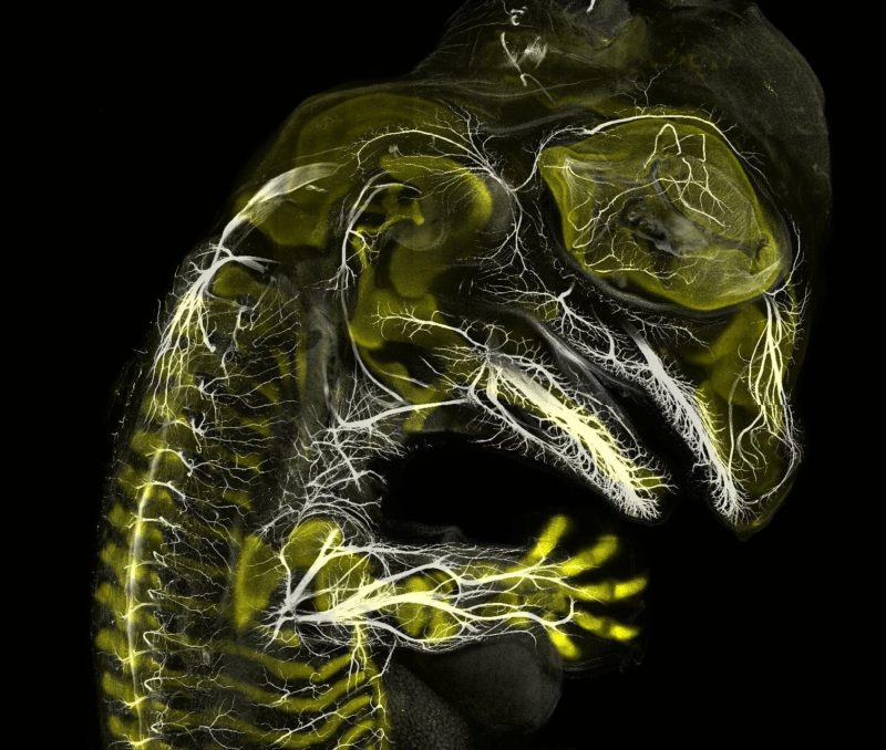
Alligator embryo developing nerves and skeleton
Daniel Smith Paredes Dr. Bhart-Anjan S. Bhullar
- Affiliation
- Yale University
Department of Geology and Geophysics
New Haven, Connecticut, USA
- Technique
- Immunofluorescence
- Magnifaction
- 10x (Objective Lens Magnification)
In Their Own Words
A Q&A with Nikon Small World winner Daniel Smith Paredes.
What is the subject matter of your winning image and why did you choose this image?
It’s an embryo of an American alligator, at around 20 days of development, stained to label the developing nerves (white) and skeleton (yellow). I thought it looked cool, and it showed how intricate the nerves can be even early in development, and how they relate spatially to the skeleton.
What are the special techniques and/or challenges faced in creating this photomicrograph?
The most difficult task was to be able to image something this size. It’s actually composed of many thousand individual pictures merged together.
What is your primary line of work?
Evolutionary developmental biology, comparative anatomy and embryology. Studying the way vertebrates have evolved by comparing their embryonic development and how it is similar and how is different between different groups of animals.
How long have you been taking photographs through a microscope?
Three-four years. I was inspired by the ability to look into the anatomical structures in developing embryos.
Do you tend to focus your microscopy toward a specific subject matter or theme? If so, why?
My interest is directed mostly towards embryonic anatomy and development.
Why did you enter the Nikon Small World Photomicrography competition? What do you think of the competition?
I was encouraged by a friend who told me I should give it a try, since my samples look good and also because they represent some groups of animals mostly understudied or photographed in micro scale.