2019 Photomicrography Competition

Fluorescent turtle embryo

Teresa Zgoda Teresa Kugler

Location
Campbell Hall, New York, USA
Technique

Stereomicroscopy, Fluorescence

Magnifaction
5x (Objective Lens Magnification)

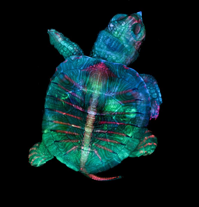
For microscopy technician Teresa Zgoda and recent university graduate Teresa Kugler, microscopy is a discipline that allows them to blend their dual passions of art and science. The 2019 winning image is a spectacular example of that, featuring a colorful turtle embryo captured using a combination of fluorescence and stereomicroscopy. 

The pair captured this image while assisting in the Marine Biological Laboratory’s embryology course. It was here they learned the precise technique required to properly prepare various types of embryos to be observed and photographed. Creating this image was a unique challenge, largely due to the size of the sample. Over an inch long, and thick, it took time and precision to ensure the entire subject was photographed completely. What’s more, the magnification used meant only a small part of the turtle could be imaged on the focal plane. The final image is a compilation of hundreds of images that had to be stacked and stitched together. 

When they are not taking photos through the microscope, both women enjoy being creative (for Kugler, that means cosplay, and for Zgoda it means photographing the landscapes, plants, and animals she sees on her hikes). Zgoda has recently started a job in a Boston hospital in a lab focused on neurology, while Kugler is excited to see what the world of science has in store for new Rochester Institute of Technology graduate. 

“Microscopy lets us get a better look at the small things in life,” said Kugler, “It allows me to do science with a purpose.” 

“We are inspired by the beautiful images we see through the microscope,” added Zgoda, “It’s amazing to be able to share that science with other people.”

In Their Own Words

A Q&A with Nikon Small World winners Teresa Zgoda and Teresa Kugler.

What is the subject matter of your winning image and why did you choose this image?

Teresa Zgoda: This image shows a turtle embryo. There aren’t many pictures of turtles that went through the skeletal prepping process, and none that were imaged using fluorescence, so we thought it would be great to share what we created.

What are the special techniques and/or challenges faced in creating this photomicrograph?

Teresa Zgoda: At the time of writing this, I have just started a new job at a research hospital in Boston where I will be assisting in a neurology microscopy core.

Teresa Kugler: I’ve recently graduated from Rochester Institute of Technology.

How long have you been taking photographs through a microscope? What first sparked your interest in photomicrography?

Teresa Zgoda: When I went to start classes at my college, I learned of the microscopy courses offered there. I remember my first microscopy class in the spring semester of my sophomore year, walking into the lab and wanting to look at every single thing under the microscope.

Teresa Kugler: Photomicrography allowed for the combination of my two favorite things, photographs and working towards a purpose in science. Just after only taking a few pictures of makeup and various plant cross-sections, I fell in love with taking pictures of the small things in life.

Do you tend to focus your microscopy toward a specific subject matter or theme? If so, why?

Teresa Zgoda: I enjoy photographing nature or natural substances, living things with cells and movement. It tends to be more interesting to me, seeing the everyday natural world at such a high magnification.

Teresa Kugler: I tend to focus on plant cross-sections because I enjoy the overall structure of the specimens.

Why did you enter the Nikon Small World Photomicrography competition? What do you think of the competition?

Teresa Zgoda: Nikon is a leading brand in the photography world, and the Small World competition brings to light a lesser known side of photography: photomicrography. The competition does wonders in curating a gallery of beautiful but also scientifically useful imagery.