Nikon Small World veteran Dr. Igor Siwanowicz took second place in the 2019 Photomicrography Competition for his composite image of three single-cell freshwater protozoans, sometimes called "trumpet animalcules.” He used confocal microscopy to capture the detail of the cilia, tiny hairs used by the animals for feeding and locomotion.
2019 Photomicrography Competition
2nd Place
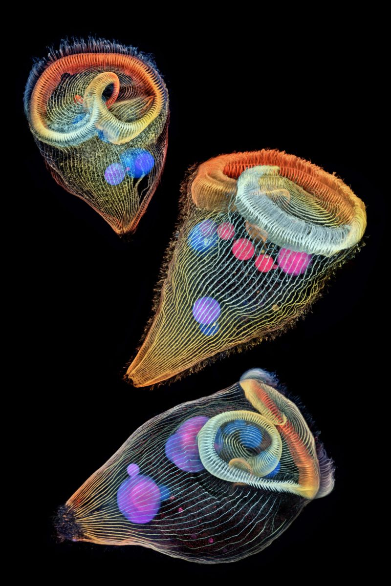
Depth-color coded projections of three stentors (single-cell freshwater protozoans)
Dr. Igor Robert Siwanowicz
- Affiliation
- Howard Hughes Medical Institute (HHMI)
Janelia Research Campus
Ashburn, Virginia, USA
- Magnifaction
- 40x (Objective Lens Magnification)
In Their Own Words
A Q&A with Nikon Small World winner Dr. Igor Siwanowicz.
What is the subject matter of your winning image and why did you choose this image?
The image, a composite of three depth color-coded projections, shows single-cell freshwater protozoans, stentors (sometimes called “trumpet animalcules”). Stentors were for quite a while on my list of potential imaging subjects, but, although present in local ponds, I could never collect a sufficient amount to try various fixation and staining protocols. Last spring, we had two Ph.D. students visiting our advanced imaging center with a goal of visualizing mitochondrial dynamics in those protozoans. I simply asked them to kindly let me use their leftover stocks after they were done with them. Stentors were featured in the Nikon Smal World contest multiple times, but I don’t recall seeing any images of the protists obtained with a confocal imaging technique. I thought that made the image somewhat unique.
What are the special techniques and/or challenges faced in creating this photomicrograph?
I used antibodies against acetylated tubulin to visualize the cilia that those tiny animals (0.5 mm long) use for feeding and locomotion, and DAPI, a DNA-binding compound, to image the nuclei that form a long strand reminiscent of a string of pearls. Fixation of the protozans is notoriously difficult. When exposed to fixative the organisms tend to collapse into a ball of protoplasm rather than retain their natural extended form. I finally found a working protocol in a 1973 publication.
What is your primary line of work?
For the past eight years, I have been studying neuroanatomy of a dragonfly, specifically the neural circuits involved in pray interception and capture. More recently, I’m collaborating on a number of projects (in- and outside of Janelia), that involve imaging of various chunks of invertebrate anatomy.
How long have you been taking photographs through a microscope? What first sparked your interest in photomicrography?
About 10 years. I was fascinated with nature since before I remember; my parents are biologists and I grew up surrounded by textbooks. I enjoyed browsing through the illustrations and photographs long before I learned how to read. It wasn’t until 16 years ago, at the age of 26, that I bought my first camera and found myself on the supply side of nature photography, with special focus on macro technique. Photomicrography is a logical continuation of photography and allows me an even more intimate perspective of my “models”.
Do you tend to focus your microscopy toward a specific subject matter or theme?
I’m enamored with invertebrate morphology; usual evolutionary restraints don’t seem to apply within the realm of tiny animals, which is evident in the abundance and variety of often grotesque and utterly alien forms. Microscopy allows me to see beyond the cuticle, explore the baroque arrangement of muscle fibers or intricate fractal-like network of neurons.
Why did you enter the Nikon Small World Photomicrography competition? What do you think of the competition?
Many scientists share an appreciation of beauty and are fully aware of the aesthetic aspects of their research. The Nikon Small World Contest was conceived with such people in mind. Images are rewarded for the artistic merit and visual aspects on par with and often above their scientific importance; that definitely grants the contest a broad appeal among non-experts and contributes to redeeming the image of science as a somber, wonder-less, unexciting affair utterly unintelligible for a layperson.